
Applications
 Breadcrumb: Home > Applications > Lab Research
 Updated:2026-11-16
 Views:
A team at the Fruit Tree Research Institute of the Guangdong Academy of Agricultural Sciences published an in planta transformation protocol for calamansi in Plants: seedlings are decapitated, Agrobacterium is applied locally to the cut surface, and about 30 days later GFP-positive shoots are screened with a LUYOR-3415RG handheld lamp using blue excitation and a yellow long-pass filter. Inoculation of 384 plants yielded 49 positive plants; for MYC2 editing, 192 inoculated plants yielded 13 edited shoots, all with mutations detected at the target site. This article sets out the workflow and the limits of fluorescence screening.
Citrus functional genomics has a persistent bottleneck: moving from a gene sequence to functional validation requires edited plants, and this step is often held up by genotype-dependent regeneration efficiency and a long juvenile phase. A recent paper in Plants offers a shorter route - no tissue culture. Agrobacterium is applied to the cut surface of a decapitated seedling, the seedling regenerates its own shoots, and fluorescence is used to pick out the positive material. The paper is specific about the lamp used for screening: a LUYOR-3415RG handheld lamp, Royal Blue excitation, with yellow long-pass filter glasses.
Citrus genome sequences are abundant, but functional validation is still held back by three things: a long juvenile phase, high heterozygosity and limited transformation capacity. Conventional Agrobacterium transformation uses epicotyl segments or other young explants, inducing regeneration in vitro and then subculturing repeatedly. Regeneration capacity varies with genotype, and the same medium may fail to produce shoots in another cultivar - so for gene function work, obtaining a batch of screenable regenerated plants takes up a great deal of time.
Reducing dependence on explant culture is the direction researchers have been pursuing. Published work has carried out in planta transformation in Shatian pomelo, Lisbon lemon, Pineapple sweet orange and rough lemon, and other routes exist such as Agrobacterium rhizogenes hairy root transformation, cut-dip-budding delivery and developmental regulator-induced meristems. What these approaches share is that transformation and regeneration are tied to the same living seedling, rather than building a regeneration system in a dish beforehand.
The recipient chosen here is calamansi (Citrus microcarpa), a hybrid of kumquat and mandarin. It has documented germplasm diversity in Hainan, polyembryonic seeds, and mature fruit available year-round under the team's production conditions - which means seedlings can be supplied continuously without seasonal constraints. The work examines material recovery and target-site editing itself; it does not assess disease resistance phenotypes.
The method: 18-20 days after germination, when the seedling has 2-4 fully expanded leaves, the shoot apex is cut off, the bacterial suspension is sprayed onto the fresh cut, and a pipette tip containing solidified medium is placed over it. The tip serves as both carrier and chamber: wide end down, the medium against the cut, film over the outside to retain moisture. This local treatment of the cut replaces in vitro culture of a whole explant.
| Step | Procedure in the paper | Key point |
|---|---|---|
| Material | Mature calamansi from a commercial orchard in Hainan, 7-13 seeds per fruit, polyembryony visible after the seed coat is removed; surface mucilage removed with 1 M sodium hydroxide for 15 minutes, sown in a peat, vermiculite, perlite 3:2:1 substrate | Fruit about 31.91 mm in transverse and 32.99 mm in longitudinal diameter, shape index 1.03; polyembryony plus a year-round fruit supply is the basis for continuous experiments |
| Culture conditions | 25±2 °C, 16 hours light / 8 hours dark, light intensity about 100 μmol m⁻² s⁻¹ | The 2-4 leaf stage is the inoculation window: too young and the seedling cannot take the cut, too old and regeneration capacity falls |
| Bacterial suspension | Agrobacterium EHA105; OD600 about 0.6 (measured on an Ultrospec 10); inoculated after treatment with 25 mg/L acetosyringone for 1-2 hours | The vector is GFP-labelled pAGM305-Cas9-gRNA-RFPA, carrying Cas9 and GFP; the empty vector simply lacks the MYC2 spacer |
| Co-cultivation | After spraying, the cut is covered with film for 45 minutes, a tip containing co-cultivation medium is fitted, and the plant is kept in the dark for 3 days; the medium contains 0.5 mg/L 6-BA, 0.1 mg/L NAA and 25 mg/L acetosyringone | The medium uses a 0.35% agarose water solution without MS salts or sucrose, supplied locally rather than by soaking the whole plant |
| Selection | Replaced with a selection tip containing 25 mg/L kanamycin, 5 days in the dark; the tip is then removed and the plant is moved to light under film | Preliminary tests showed that 50 mg/L clearly inhibited shoot regeneration, so the selection pressure was set at 25 mg/L |
| Observation | GFP observation about 30 days after inoculation, with continued monitoring during later growth | Counting from germination, an initial fluorescence read is possible in about 7 weeks |
Antibiotic selection can tell you whether a seedling has been killed by the selection agent, but not whether the T-DNA is being expressed. Confirming expression takes either a reporter gene or molecular tests such as PCR and sequencing - the latter require sampling, DNA extraction and waiting for results, are costly, and sampling itself may damage a newly regenerated shoot. The paper therefore works in the order in situ fluorescence screening to narrow the candidates, then molecular testing for the qualitative result.
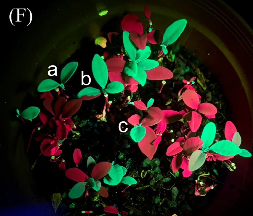
The paper records the equipment and parameters for this step clearly: a LUYOR-3415RG handheld lamp, Royal Blue excitation (specified 440-460 nm), with yellow barrier glasses (specified 500 nm long-pass cut-off); images were taken with an iPhone 16 Pro Max camera through the same filter. This configuration matches the RB channel of the 3415RG: blue light excites GFP, the filter glasses block the excitation light, and the YELLOW long-pass passes the green fluorescent signal while blocking the rest.
| Operating point | Procedure in the paper | Why it is done this way |
|---|---|---|
| Observation timing | About 30 days after inoculation, once regenerated tissue has formed | Too early and the signal is insufficient and confused with selection death; too late and positive shoots have grown into the same mass as negative ones |
| Excitation and filtering | RB 440-460 nm excitation, yellow 500 nm long-pass filter glasses | The EGFP excitation peak is near 488 nm, so blue light is enough to excite it; the long-pass filter removes excitation light and part of the background, so the green signal can be read |
| Imaging | Phone camera placed behind the filter, paired with a brightfield image of the same field | Fluorescence tells you this area is bright; the brightfield image locates which shoot and which leaf |
| Result categories | Uniformly GFP-positive shoots, chimeric shoots and single-leaf fluorescence counted separately | Different distributions imply different transformation timing and degrees of chimerism, and later sampling strategies differ |
| Interpreting negatives | Regenerated shoots treated identically but not transformed served as controls; their red fluorescence was judged to be chlorophyll autofluorescence, not RFP expression | Plant tissue has red autofluorescence of its own; without a control the background is easily read as positive |
Separating the red and green signals is central to this reading. The paper states explicitly that GFP-negative control plants also showed red fluorescence, interpreted as chlorophyll-related autofluorescence rather than reporter RFP expression. Green fluorescence that could be reproduced was counted as GFP-positive. The experience is general in plant fluorescence observation: establish what counts as bright against a negative background before discussing a positive call.
The empty-vector experiment ran four batches of 96 plants each, using a vector that carries GFP but no MYC2 spacer, specifically to measure how many inoculated plants produce GFP-positive tissue:
| Batch | Plants inoculated | Uniform positive shoots | Chimeric shoots | Single-leaf fluorescence | Total GFP-positive | Positive rate |
|---|---|---|---|---|---|---|
| B1 | 96 | 2 | 1 | 2 | 5 | 5.2% |
| B2 | 96 | 10 | 2 | 2 | 14 | 14.6% |
| B3 | 96 | 13 | 3 | 2 | 18 | 18.8% |
| B4 | 96 | 8 | 2 | 2 | 12 | 12.5% |
| Total | 384 | 33 | 8 | 8 | 49 | 12.8% |
Positive rates across the four batches ranged from 5.2% to 18.8%, mean ± standard deviation 12.8±5.7%; 33 plants produced uniformly GFP-positive shoots, 8.6% of inoculated plants. This indicator counts plants - a plant with several positive shoots counts once - so it measures recovery of positive tissue, not shoot regeneration rate.
The editing experiment was a separate batch: 192 plants were inoculated with the vector carrying the MYC2-targeting spacer, and 13 GFP-positive shoots (L1-L13) were recovered, a positive rate of 6.8%. All 13 amplified a GFP-specific band, and amplicon sequencing of the target site detected mutations in all 13; the edited read proportion in individual samples ranged from 82.8% to 99.5%, with most samples showing predominantly insertions and L9 predominantly deletions. The target lies at the 5′ end of the MYC2 coding region, the spacer is 5′-GGATTGCCAGAATATGGCGT-3′, and the adjacent PAM is 5′-AGG-3′. MYC2 encodes a bHLH transcription factor, and published work indicates that stabilising this protein can enhance citrus resistance to huanglongbing; this study verifies material recovery and target-site editing, and did not test disease resistance phenotypes.
The team also ran a single preliminary batch on Shatian pomelo: 96 seedlings were inoculated with a GFP-labelled overexpression vector, 6 showed GFP-positive tissue (6.25%), and all 6 gave the expected fragment in GFP-specific PCR; negative control plants showed red chlorophyll autofluorescence alone. The authors note that both the recipient material and the vector differ from the calamansi experiment, so the two frequencies cannot be compared directly; this step is a proof of concept.
Fluorescence is a preliminary screen, not a conclusion. The handheld lamp answers whether this shoot is glowing and worth sampling; PCR and sequencing answer whether the T-DNA integrated and the target site changed. In the paper all 13 positive shoots went through GFP-specific PCR and target-site sequencing, which is exactly this division of labour: the lamp narrows the candidates to a dozen or so plants, and molecular testing gives the qualitative result.
Fluorescence distribution carries more information than its mere presence. Uniformly positive shoots, chimeric shoots and single-leaf fluorescence were recorded separately: uniform fluorescence means transformation happened early and the regenerated shoot derives from transformed cells, while chimerism and single-leaf fluorescence often indicate transformation in tissue that had already layered, so later work has to watch whether the genotype is consistent. Choosing uniformly fluorescent shoots at sampling time can save a great deal of later verification.
Signal intensity changes, so the criterion has to be stable. The paper records that representative regenerated tissue still carried GFP fluorescence about a year after inoculation, which shows positive material can be followed over time; it also states that fluorescence positivity is not the same as genetic stability, and that integration status, heritability and uniformity still need further characterisation. Ageing plant tissue and reporter gene silencing both lower the signal, so weak fluorescence cannot be judged negative outright.
LUYOR-3415RG dual-wavelength fluorescent protein excitation source - RB for GFP, GR for DsRed, handheld observation without a darkroom; EGFP screening in alfalfa shoot-tip transformation: a LUYOR-3415RG application - another fluorescence screening case in a plant regeneration system; GFP screening in grape hairy root transformation - fluorescence validation across 12 genotypes; GFP fluorescence flashlight section to see more plant fluorescence observation options, or contact us for 3415RG selection advice and a trial.

Email: info@xinysun.com

Tel: 131-2093-6518

Address: 1525 Minqiang Road, Bldg 26, Songjiang, Shanghai

Copyright 2026 Xinysun Instrument (Shanghai) Co., Ltd. All rights reserved.
ICP:ICP-2026046447




 Industrial Test Instruments


