
Products
PRODUCTSGFP excitation flashlights use a specific wavelength with a filter for observing green fluorescent protein (GFP) and other fluorescent labels in molecular biology and life science experiments, aiding fluorescence imaging and teaching demos. Portable and easy to use, they enable rapid screening of fluorescence expression in a darkroom or on site, with excitation wavelengths and observation filters available by experiment. In stock in Shanghai with selection consulting.
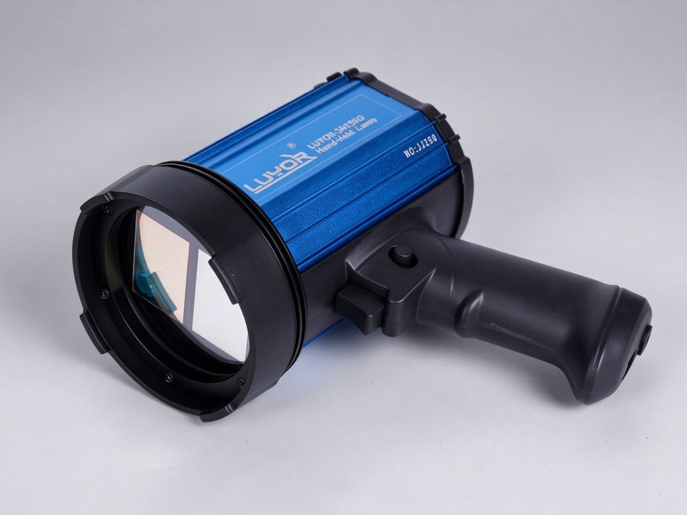
 Model: LUYOR-3415RG

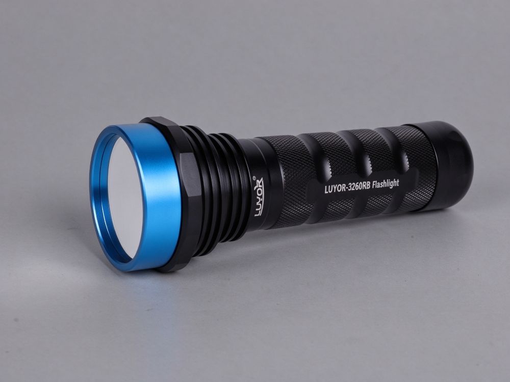
Model: LUYOR-3260RB/3260GR
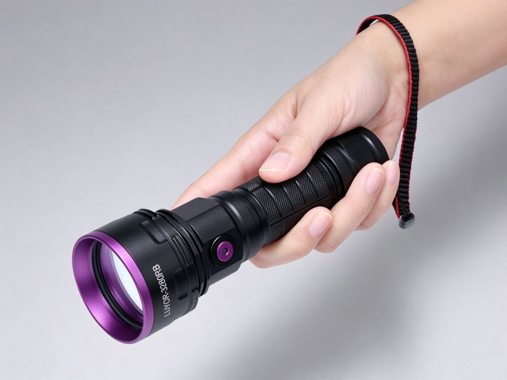
Model: LUYOR-3280RB/3280GR
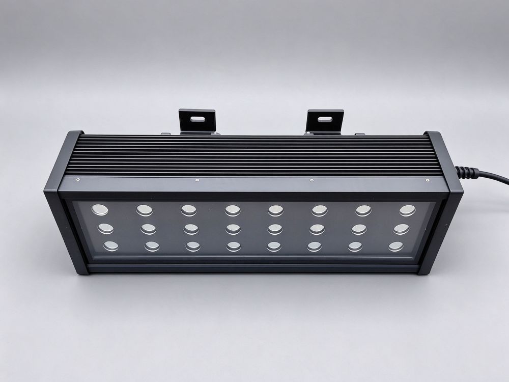
Model: LUYOR-3124RB
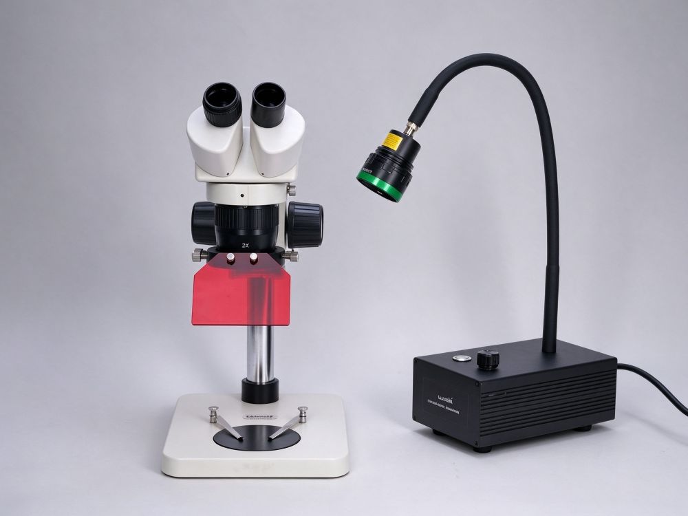
Model: LUYOR-3421RB/LUYOR-3421GR
Select an excitation light source by starting with the wavelength: GFP and eGFP have excitation peaks near 488nm and require 440-460nm blue light plus 500nm long-pass filter goggles; DsRed and mCherry excite near 550nm and need 510-540nm green light. Once the wavelength is settled, compare coverage area and battery life: whole trays of plants or groups of mice need a large area, while single-plant screening and field observation favor portability.
| Model | Excitation Band | Coverage Area | Power / Supply | Typical Applications |
|---|---|---|---|---|
| LUYOR-3415RG | Dual wavelength selectable: RB 440-460nm / GR 510-540nm | Ø180mm at 150mm | 27W, 0.9kg total weight | One unit handles both GFP and DsRed |
| LUYOR-3260RB | 450nm deep blue | Ø150mm at 30cm | 9×3W, 12V lithium battery, 2-4 hours | Handheld screening, seed sorting |
| LUYOR-3280RB/GR | Over 10 selectable bands | Ø300mm at 50cm | 20W, dual batteries 4 hours, Type-C | Whole-plant and whole-tray observation and imaging |
| LUYOR-3124RB | 440-460nm narrow band | 180×450mm at 40cm | 24 high-power LEDs, hanging mount | Whole trays of plants, groups of mice |
| LUYOR-3421 | Quick-swap modules: 365/405/450/488/505/530nm | Large-area uniform illumination | 20W benchtop, BNC quick-swap | Microscope adapter, fruit fly and zebrafish screening |
| NIGHTSEA Xite-RB / GR | RB 440-460nm / GR 510-540nm | Handheld illumination | 255g, two power levels + strobe mode | Portable field and on-site observation |
The comparisons above are compiled from publicly available parameters on the individual product pages. For exact configuration, pricing and lead time, please refer to the corresponding product page or call the number at the bottom of this page. Xinysun Instruments (Shanghai) Co., Ltd. supplies LUYOR and XEPU instruments for industrial inspection, UV curing and speed measurement.
Fluorescent proteins such as GFP and eGFP absorb energy under light of a specific wavelength and re-emit it as green fluorescence at a longer wavelength; DsRed and mCherry absorb blue-green light and emit orange-red. The excitation light source concentrates energy at the protein's absorption peak, while matched long-pass filter goggles block the excitation light and let only the fluorescence reach the eye or camera — so fluorescence equals expression.
It is therefore never an ordinary flashlight: the wavelength must match the protein's absorption peak and the goggles must block the excitation band; miss either one and you will see either a wash of blue light or nothing at all. Choose in this order: protein first, then wavelength and coverage area, and only then brightness and battery life.
1. Match protein to wavelength: Use 440-460nm blue light with a 500nm long-pass for GFP/eGFP; use 510-540nm green light with the corresponding long-pass for DsRed/mCherry.
2. Dim the environment: It need not be pitch dark, but lower ambient light gives a lower background; use a hood for quantification or imaging.
3. Wear matched goggles: Put on the goggles before switching on the lamp to avoid looking directly at the excitation light; use a 500nm long-pass for blue excitation and the corresponding blocking filter for green excitation.
4. Control distance and angle: Irradiate at the stated distance (commonly 15-30cm), with the spot just covering the target; a slight off-axis angle reduces specular reflection.
5. Interpret and document: Positive tissue/plants show bright green (or red) fluorescence; recheck weak signals from another angle. When photographing, lock white balance and exposure and use the same settings for the whole batch.
Choose the band by protein first, then the form factor by sample scale: handheld for a single plant or dish, hanging for whole trays or cages, benchtop for microscopes and well plates.
| Observation Target | Excitation Band | Typical Form Factor | Notes |
|---|---|---|---|
| GFP / eGFP | 440-460nm blue light + 500nm long-pass | Handheld or hanging | The most widely used combination, suitable for both plants and zebrafish |
| DsRed / mCherry | 510-540nm green light + long-pass | Switch on a dual-wavelength model | Screening for GFP and DsRed together requires a dual-wavelength model or two units |
| Whole trays of plants, groups of mice | Narrow band 440-460nm | Hanging type, covering the tray | Coverage area takes priority over portability |
| Well plates and microscopes | 365/405/450/488/530nm modules | Benchtop, quick-swap modules | Choose the module according to the dye and filter combination |
- Plant transformation screening: Preliminary screening of positive T1 seeds, seedlings and callus tissue.
- Zebrafish and fruit flies: Fluorescent labeling of embryos, observation of nerves and blood vessels.
- In vivo mouse imaging: Preliminary fluorescence screening of ears, tails and subcutaneous xenografts.
- Cells and organoids: Rapid assessment of transfection efficiency in well plates and dishes.
- Microorganisms and yeast: Colony screening with fluorescent reporters.
- Teaching and outreach: Fluorescent protein demonstrations — visual and safe.
- Selection and Brands: Avoiding Pitfalls in Choosing a Fluorescent Protein Excitation Source: Why Do So Many Experiments Show No Fluorescence?, How to Choose a UV Crosslinker vs. a Fluorescent Protein Excitation Light Source? A Guide to UV and Fluorescence Tools for Life Science
- Operation and Interpretation: Hairy Root Transformation of Raspberry Without Tissue Culture: How to Rapidly Screen eGFP-Positive Roots, Colony Counting in Bacterial Competition Assays: How to Perform mCherry Fluorescence Sorting, Preliminary GFP Fluorescence Screening of Transgenic Adventitious Roots: How to Use a 365nm Blacklight
It depends on band coverage. GFP and eGFP have excitation peaks near 488nm and are viewed with 440-460nm blue light plus 500nm long-pass filter goggles; DsRed and mCherry excite near 550nm and require 510-540nm green light. A dual-wavelength model such as the LUYOR-3415RG switches between the two bands, covering both in one unit; a single-wavelength model handles only one protein, so decide which you need before choosing.
Not mandatory. Most models (such as the 3415RG and 3280) are rated for observation without a darkroom: simply wear matched long-pass filter goggles and dim the ambient light enough to work comfortably. For fluorescence quantification or photographic documentation, a darkroom or hood is still recommended to prevent ambient light from raising the background and lowering the signal-to-noise ratio.
Excitation light is far more intense than the fluorescence signal, so looking directly is overwhelming — you see no fluorescence at all, and prolonged exposure poses a safety risk. Filter goggles block the excitation band and pass only the fluorescence, creating the contrast of a dark background with a bright green signal. The goggles must match the excitation band: a 500nm long-pass for blue excitation and the corresponding blocking filter for green excitation.
It can show both, but not simultaneously. A dual-wavelength model switches between two bands: blue light for GFP, then switch to green for DsRed. To image both colors in the same sample at once you need simultaneous dual-channel output with a dual-band filter, which belongs to imaging systems and is beyond a handheld lamp.
Four approaches: reduce ambient light (use a hood), shorten the irradiation distance to concentrate the spot, switch to higher power or a narrower band, and confirm that the goggles match the band (a mismatch filters out the fluorescence too). If the sample itself expresses at low levels, no light source can rescue it — go back and optimize the vector and promoter.
Yes, but turn off auto white balance and auto exposure, manually reduce exposure by one or two stops, and hold a goggle lens in front of the camera as a filter. Keep ambient light as low as possible when photographing GFP, or the green will be washed out by white light; publication-quality images still require an imaging system with a filter wheel.
Brief observation has little effect, but prolonged high-intensity illumination can cause phototoxicity and thermal effects, and plant leaves may show localized bleaching. Limit irradiation at any one spot to a few minutes; for long-term observation reduce the power or use a pulsed/strobe mode.

Email: info@xinysun.com

Tel: 131-2093-6518

Address: 1525 Minqiang Road, Bldg 26, Songjiang, Shanghai

Copyright 2026 Xinysun Instrument (Shanghai) Co., Ltd. All rights reserved.
ICP:ICP-2026046447




 Breadcrumb:
 Products
 Applications
 Industrial Test Instruments


