
Applications
 Breadcrumb: Home > Applications > Lab Research
 Updated:2026-11-06
 Views:
This article takes apart the three-layer design of a green Sichuan pepper transformation platform: how the regeneration system brings the shoot-to-plantlet cycle under two months, how the RUBY visible marker gives a readout for four transformation parameters - Agrobacterium concentration, infection time, dark co-cultivation and cefotaxime level - and why DsRed2-positive material is then handed to a portable fluorescence lamp for in situ prescreening 40 days after infection. It also sets out the waveband and filter glasses the 3415 series uses to observe red fluorescence, and where the boundary lies between fluorescence prescreening and molecular verification such as PCR and RT-qPCR.
Green Sichuan pepper is a perennial deciduous shrub in the genus Zanthoxylum, family Rutaceae, valued both as a medicinal and as a food plant, and concentrated in southwest China. The paper records that annual Chinese pepper output has exceeded 300,000 tonnes for several years running, with an industry worth billions of US dollars. Over the past few years a good deal of genomic, transcriptomic and metabolomic data has accumulated for the species, but functional genomics has not moved forward - without an efficient regeneration and genetic transformation system, candidate genes cannot be verified in the plant itself.
Tissue culture of woody economic species is difficult, and the difficulty usually lies in the material. Green Sichuan pepper is rich in secondary metabolites such as alkaloids, terpenes, flavonoids and coumarins, so explants brown and necrose readily once excised; field-collected material also carries a heavy load of endophytes. This study switched to stem segments from one-year-old seedling plants raised in a greenhouse, which reduces browning and lowers the initial microbial load, laying a base for the two-step workflow that follows.
Transformation has to land on tissue that already regenerates, so regeneration efficiency has to be stable to begin with. The study split the workflow into four stages - disinfection, shoot induction, rooting and acclimatization - and fixed the parameters for each.
10 minutes of ultrasonic pretreatment, followed by 75% ethanol for 45 s and 5% NaClO for 8 minutes. With chemical disinfection alone, 8 minutes of 5% NaClO gave a contamination rate of 27.01%, which fell to 21.33% with ultrasound; extending ultrasound to 15 minutes caused obvious browning and raised the death rate.
With MS as the basal medium, nine combinations of 6-BA and IBA were compared. A9 (1.5 mg/L 6-BA + 0.2 mg/L IBA) gave 88.33% shoot formation at 14 days, significantly above A1 without hormones (about 30%) and A7 with IBA alone (65.01%).
1/4 MS with 0.5 mg/L IBA (B2) stood out: 85.01% rooting, mean root length 4.77 cm, mean time to rooting 30.01 days; raising IBA to 1.5 mg/L brought rooting back down to 30.00%.
After 35 days of rooting culture, lids were loosened gradually over 3-5 days, medium was washed from the roots, leaves were removed, and plantlets were moved to a 1:2 peat and vermiculite substrate under film to hold humidity. After 60 days survival was about 93.33%, with mean height up 7 cm and stem diameter up 0.4 cm.
The authors also reran the workflow with three seedling individuals of the same cultivar (G1, G2, G3), obtaining 73.00%-83.67% shoot formation and 67.67%-85.12% rooting, which shows the parameters are not effective for a single genotype alone. The three genotypes differed, but all of them worked, which matters for functional experiments that have to be repeated across materials.
| Stage | Key conditions | Result |
|---|---|---|
| Disinfection | Ultrasound 10 min + 75% ethanol 45 s + 5% NaClO 8 min | Contamination rate 21.33% |
| Shoot induction (A9) | MS + 1.5 mg/L 6-BA + 0.2 mg/L IBA, 14 days | Shoot formation 88.33% |
| Rooting (B2) | 1/4 MS + 0.5 mg/L IBA | Rooting 85.01%, 30.01 days, root length 4.77 cm |
| Acclimatization | 35-day rooted plantlets, peat:vermiculite 1:2, 60 days | Survival 93.33% |
Once the regeneration system was stable, optimizing transformation parameters still needed a reliable readout. The study chose RUBY - a visible reporter gene that makes plant tissue accumulate betalains and turn red to the naked eye. It needs no excitation light and no sampling; positive explants can be counted in the dish. Defining the RUBY-positive transformation frequency as red-marked explants divided by total infected explants, four parameters were scanned one at a time:
| Parameter | Gradient and readings | Conclusion |
|---|---|---|
| Agrobacterium concentration, OD600 | 0.4 gave 14.33%, 0.6 rose to 36.01%, and further increases lowered both expression and explant vigor | 0.6 |
| Infection time | 20 min gave 22.83%, 30 min rose to 38.47%, at 40 min Agrobacterium overgrowth appeared | 30 min |
| Dark co-cultivation | 12 h gave just 11.91%, 24 h reached 32.60%, at 48 h microbial overgrowth killed the tissue | 24 h |
| Cefotaxime | 0 mg/L gave 6.60%, 300 mg/L reached 56.37%, 400 mg/L gave 44.93% with suppressed explant development | 300 mg/L |
Putting the four together, the recommended combination is OD600 = 0.6, 30 minutes of infection, 24 hours of dark co-cultivation and 300 mg/L cefotaxime in the medium, at which the RUBY-positive transformation frequency reached the study's peak of 56.37%, with clear red visible on both stem segments and newly formed organs. To confirm the red was not an artefact, the authors randomly picked 5 RUBY-positive materials, extracted genomic DNA and amplified a 500 bp fragment of the RUBY sequence; all five samples gave a band and the wild-type control gave none.
Reading boundary: 56.37% is the "RUBY-positive transformation frequency", measuring the proportion of explants carrying a visible marker 40 days after infection. It is not the same as a stable, heritable transformation efficiency. The paper itself positions it as a prescreening indicator in the discussion, with molecular testing and progeny verification still to follow.
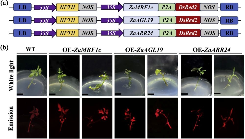
RUBY is good for optimizing parameters in batches, but verifying a functional gene needs a different readout. The study chose three target genes with existing research foundations - ZaMBF1c, ZaAGL19 and ZaARR24 - driven by the 35S promoter, with the target protein fused to the red fluorescent protein DsRed2 through the P2A self-cleaving peptide, so that both proteins are expressed at once without interfering with the function of the target gene. The vector backbone carries NPTII kanamycin resistance.
The paper states that 40 days after infection, a portable fluorescence lamp LUYOR-3415 was used to identify reporter-positive individuals efficiently. In the results, DsRed2-positive material grew in the same way as wild type under natural conditions, and red fluorescence was detected in positive material, while WT controls did not light up. The value of this step is that it is non-destructive, in situ and immediate: the whole batch of regenerated plantlets is screened once, candidates are picked out, and PCR and RT-qPCR are left to verify a small number of samples.
The verification chain was also fairly complete. Before sampling, positive plantlets were washed three times for 3 minutes each with sterile water containing 300 mg/L cefotaxime and then rinsed three times with sterile water, to remove residual Agrobacterium on the tissue surface that would interfere with nucleic acid extraction; material infected with the empty-vector GV3101 served as a negative control. PCR then amplified target bands of the expected size, and RT-qPCR (ZaUBQ as the internal reference, 2^-ΔΔCt method) showed the three genes upregulated 2.56-5.38 fold relative to wild type, with 3 independent positive materials and 3 technical replicates per gene.
"...facilitating efficient identification of reporter-positive individuals after infected 40 days using a portable fluorescence lamp LUYOR-3415". The paper records the model LUYOR-3415 and the observation time point (40 days after infection), and does not state the waveband, filter glasses or illumination distance; the configuration below is product-side information, for reference when reproducing the experiment.
DsRed2 is a red fluorescent protein with its excitation peak in the green region, so what it needs is not the familiar royal-blue GFP excitation but the GR green band. Among the light source options for the LUYOR 3415 series, the match is GR (510-540 nm), with LUV-50A barrier filter glasses on the observation side (590 nm long-pass, for DsRed, tdTomato, mCherry and other RFP-family proteins); if GFP or eGFP also has to be observed, switch to RB royal blue 440-460 nm and change to LUV-30A glasses.
The corresponding model sold through our site is the LUYOR-3415RG dual-wavelength fluorescent protein excitation source, which carries both the RB and GR bands in one unit with two-switch selection, covering GFP and DsRed labels. Key specifications: 27 W total (6 x 3 W LED per band, 12 in total), net weight 0.9 kg, 180 mm illumination diameter at 150 mm from the lamp, rechargeable battery power, no darkroom required, a light-proof cloth included, and an optional adjustable stand and photographic filter.
| Target | Excitation band | Viewing glasses | Typical use |
|---|---|---|---|
| GFP, eGFP | RB royal blue 440-460 nm | LUV-30A | Green-fluorescent seedlings, callus and roots |
| DsRed2, mCherry, tdTomato | GR green 510-540 nm | LUV-50A | Red-fluorescent positive material, seed sorting |
| GFP in green tissue | RB royal blue 440-460 nm | LUV-30AG | Suppressing the red chlorophyll autofluorescence background |
Placed in this green Sichuan pepper workflow, three points about use are worth noting. To start with, a 180 mm spot at 150 mm is enough to illuminate a whole regenerated plantlet in one go, with no need to move the lamp back and forth, and culture vessels can be illuminated through the wall, so no contamination is introduced by opening them. Second, at day 40 a whole batch has to be processed, so fixing the lamp on the optional stand and freeing both hands for picking saves time compared with holding it. Third, when images are needed for records, fit the photographic filter, lock exposure and white balance manually, and turn the flash off.
If positive tissue later has to go under a stereomicroscope to see where expression is located, the LUYOR-3421 benchtop fluorescent protein excitation source can be used instead: it has six quick-change modules at 365/405/450/488/505/530 nm, works as a microscope fluorescence adapter and can also illuminate plates on its own; for large-area whole-plantlet prescreening the handheld 3415 series is still easier to handle.
Judgment reminder: a fluorescence-positive result is a candidate, not proof of stable transformation; for weakly expressing material, observe in shade or with the light excluded, since chlorophyll autofluorescence in green tissue raises the background in the red channel; residual Agrobacterium and medium components can also interfere, which is why the paper arranged cefotaxime washing and a negative control. The conclusion holds once the 3415 reading is chained to PCR, RT-qPCR and progeny segregation analysis.
What is really reusable is not a particular medium recipe but the split into two stages: sweep the infection conditions with the naked-eye RUBY marker and fix the parameters, then switch to a fluorescent reporter for formal functional verification, using a portable excitation source for non-destructive prescreening and concentrating expensive molecular testing on the candidates. For woody and economic trees that regenerate slowly, brown heavily and are expensive in material, this order saves a great deal of tissue culture and sequencing work.
Comparable cases on this site include EGFP prescreening in alfalfa shoot-tip transformation, fluorescence reading in grape hairy root transformation and visual reporters for in vivo transformation in macadamia. All follow the same logic - a visible marker to fix parameters, a fluorescence source to pick positives, molecular testing to close the loop - and differ in little more than the color of the reporter and the sampling window.
The two do different jobs. RUBY turns tissue red directly, so it can be counted by eye with no equipment, which suits rapid comparison of a dozen or so treatments in OD600, infection time, co-cultivation time and antibiotic concentration during parameter optimization; DsRed2 is a fluorescent protein co-expressed with the target gene through P2A, used to judge whether the target gene has actually been delivered and is being expressed, and it needs an excitation source and filter glasses. The former fixes the conditions, the latter verifies the function.
3415 is the series name of the LUYOR portable fluorescent protein excitation source, and the paper records the model at series level without specifying the band configuration; 3415RG is the corresponding dual-wavelength configuration sold through our site, carrying RB (440-460 nm) and GR (510-540 nm) light sources. Observing DsRed2 red fluorescence uses the GR band with LUV-50A glasses. The paper does not state the band, so when reproducing the work the configuration should follow the excitation requirement of the reporter.
Excite with GR green at 510-540 nm and observe through LUV-50A barrier filter glasses (590 nm long-pass), so that red fluorescence separates from the reflected excitation light. If the material is also labeled with GFP, switch to RB royal blue 440-460 nm and change to LUV-30A. Glasses cannot be mixed, otherwise background light will swamp the fluorescence signal.
Yes. Prescreening answers "this plant expresses the reporter" and cannot answer whether the T-DNA is stably integrated, how many copies there are, or what the transcript level of the target gene is. After picking DsRed2-positive material, the paper still ran PCR for the target fragment, RT-qPCR with ZaUBQ as the internal reference, and a negative control infected with empty-vector GV3101, obtaining 2.56-5.38 fold upregulation of the three genes before the verification loop was considered closed.
The 3415 series is handheld, and its 27 W output is enough to observe under ordinary indoor light, so no darkroom has to be set aside; in a greenhouse or field with strong midday sun contrast will fall, and shade or the supplied light-proof cloth is advisable. Among the customer practice recorded on the product page, one research institute illuminated culture plantlets directly through the vessel wall to judge positives, which avoids contamination by not opening the vessel. For batch screening an adjustable stand is advisable, and for images the photographic filter.
No. That figure is the RUBY-positive transformation frequency under 300 mg/L cefotaxime, that is, the proportion of explants carrying a visible red marker 40 days after infection, and it is a prescreening reading. Stable transformation still requires molecular testing, confirmation of regenerated plants and progeny segregation analysis. It is more accurate to read it as an upper reading from parameter optimization.
Qi Z, Luo Z, He Y, Chen Y, Huang Z, Wang J, Wang L, Xiong Y, Cui B, Gong W, Hui W. A rapid regeneration and visible-marker-assisted transformation platform supporting gene function studies in Zanthoxylum armatum. Scientia Horticulturae, 2026, 369: 115291. doi:10.1016/j.scienta.2026.115291 (online 2 November 2026, CC BY-NC). The equipment is recorded in the paper as a portable fluorescence lamp LUYOR-3415.
Related products: LUYOR-3415RG dual-wavelength fluorescent protein excitation source, LUYOR-3421 benchtop fluorescent protein excitation source, LUYOR-3260RB fluorescent protein excitation source. Xinyisheng Instruments (Shanghai) supplies the full LUYOR range of fluorescence observation equipment and can help match waveband, filter glasses and stand to the reporter.
This article belongs to the laboratory research techniques section.
Other sections on the same product line include GFP fluorescence flashlights (models sorted by GFP/RFP/DsRed band, handheld and benchtop) and life science instruments (the entry point for fluorescence imaging, UV crosslinking, UV transmission and cell irradiation).
Models mentioned in this article: LUYOR-3415, LUYOR-3421, LUYOR-3260RB. Ranges, accuracy and accessory lists for each model are given on the product pages.

Email: info@xinysun.com

Tel: 131-2093-6518

Address: 1525 Minqiang Road, Bldg 26, Songjiang, Shanghai

Copyright 2026 Xinysun Instrument (Shanghai) Co., Ltd. All rights reserved.
ICP:ICP-2026046447




 Industrial Test Instruments


