
Applications
 Breadcrumb: Home > Applications > Lab Research
 Updated:2026-11-16
 Views:
Plant breeding, model-organism work and microbial labeling share three common bottlenecks: scarce fluorescence microscope time, inconvenient field sampling, and the cost of PCR-verifying large batches. This article presents the LUYOR-3415 portable dual-wavelength excitation source as a pre-screening tool ahead of the microscope. It supports non-destructive in situ viewing in greenhouses, fields and animal rooms, and allows rapid initial sorting of GFP, DsRed, CFP and DAPI labeled material. The RG, RC, RV and RU wavelength combinations are listed with their applications, followed by three scenarios, a four-step workflow and the precautions that keep the method reliable. The instrument is positioned for visual high-throughput screening and does not replace high-resolution fluorescence imaging.
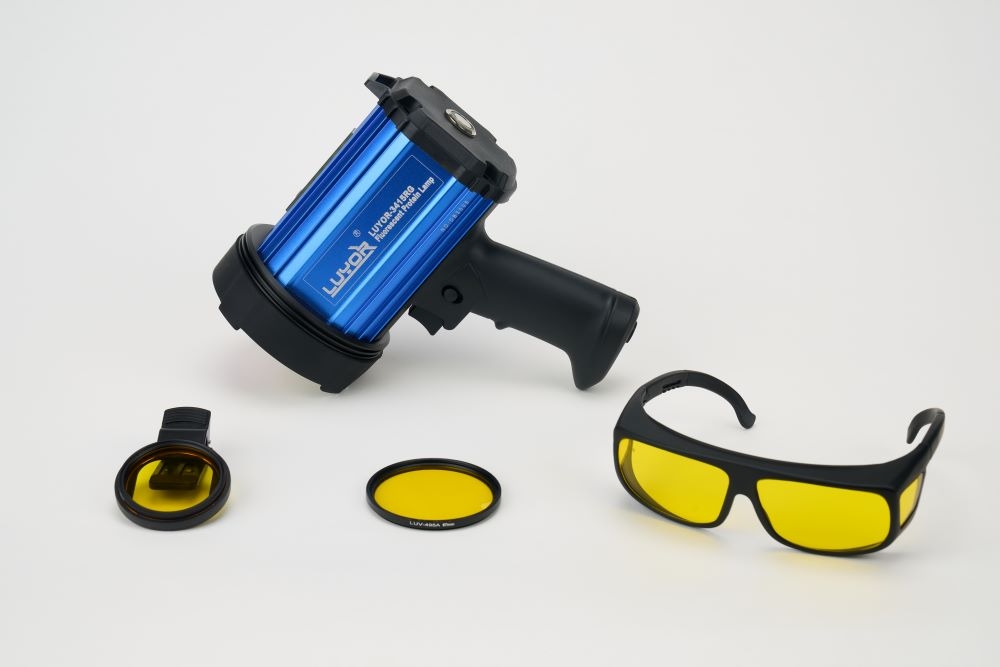
In plant breeding, model-organism and microbial fluorescent labeling work, three problems are common: fluorescence microscope time is scarce, in situ sampling in the field is inconvenient, and PCR validation of large batches is expensive. The LUYOR-3415 portable dual-wavelength fluorescent protein excitation source is designed as a pre-screening tool that works alongside the microscope. It supports non-destructive in situ viewing in greenhouses, fields and animal rooms, and allows rapid initial sorting of GFP, DsRed, CFP and DAPI labeled material, which in most cases reduces experimental cost and raises screening throughput.
Transgenic function validation, screening of gene-edited material and observation of fluorescently labeled model organisms normally depend on fluorescence microscope imaging. When sample numbers are large, mounting each sample and booking instrument time consumes a great deal of capacity; if every sample goes to nucleic acid testing, reagent and labor costs are high and the cycle is long. Many positive plants can be recognized by eye, without immediate subcellular imaging. That is the basis for an in situ pre-screening step: a portable excitation source removes negative individuals quickly, so that suspected positives go on to PCR, sequencing or microscope confirmation.
Note on positioning: the LUYOR-3415 is intended for visual high-throughput pre-screening and does not replace high-resolution fluorescence microscopy.
The LUYOR-3415 uses a high-power narrow-band LED source with two selectable excitation wavelengths, switched with one button. The unit runs on a rechargeable lithium battery and can be hand-held, or mounted on a stand for long sessions.
Selectable wavelength pairs for different fluorescent labels
RG (450nm+520nm): GFP/eGFP and DsRed, the usual pairing in breeding work
RC (405nm+450nm): CFP and GFP
RV (405nm+520nm): CFP and DsRed
RU (365nm+450nm): DAPI and GFP
An even spot for large trays and seed batches Unlike ordinary fluorescence torches, which fall off sharply at the edge, a large even spot lowers the chance of missing weak fluorescence. With the supplied light-shielding cloth no darkroom is needed, so observation can take place in a greenhouse, a growth room or a field at dusk.
Non-destructive viewing that leaves the sample intact No leaf clipping and no sampling are needed; positive seedlings can be transplanted or subcultured directly. The method suits seedling-stage and hairy-root observation in Arabidopsis, rice, maize, soybean and cotton, and live observation of model animals such as zebrafish and mice.
Matched filter glasses They block stray excitation light, raise fluorescence contrast and protect the eyes. Screening records can be photographed with a phone and kept in the laboratory log.
Material: transformed seedlings, hairy roots and T1 seeds of Arabidopsis, rice, maize, soybean, peanut, tobacco, cotton and potato. Workflow:
After the seedling stage or hairy-root induction, place the plants under dim light and lay out the light-shielding cloth;
Select the matching wavelength channel, switch on the source and observe through the filter glasses;
Mark fluorescent-positive individuals and discard negative plants directly;
Sample the positive material for PCR validation, or send it for detailed microscope imaging. Cases: agricultural and forestry universities, academies of agricultural sciences and commercial seed companies use this model for soybean hairy-root work, maize seed, potato edited mutants and peanut rhizobium colonization screening; overseas seed company GDM uses it for large-batch transgenic seed sorting.
Zebrafish embryos, fluorescently labeled mouse tissue and nematode fluorescent reporter strains. Positive and negative individuals can be separated in situ in the animal room, which reduces anesthesia and dissection sampling.
Observation of fluorescently labeled rhizobia, engineered bacteria and fungal colonies on plates, for rapid picking of positive colonies.
Confirm the fluorescent protein carried by the construct before choosing a wavelength; a mismatch makes fluorescence invisible;
Strong midday sunlight drowns the signal, so work at dusk, in a shaded greenhouse or in a growth room;
Never point the excitation light at the eyes; wear the matched filter glasses during observation;
Reserve the instrument for visual pre-screening and records; high-resolution publication images still require a fluorescence microscope.
Lower cost: far fewer samples go to DNA extraction, PCR and sequencing, which saves reagents;
Freed instrument time: less low-value screening on the microscope, more capacity for detailed imaging;
Higher throughput: screening happens in the field or greenhouse, with no samples carried back to the laboratory;
Flexible deployment: battery operation suits research groups, shared platforms and seed companies alike.
The model is cited in publications from universities, research institutes and seed companies in China, and a deposit-based demo unit with full selection guidance is available to research groups.
This article belongs to the Research and Laboratory Techniques section.
Other sections on the same product line include GFP Flashlights (models grouped by GFP/RFP/DsRed band, hand-held and benchtop) and Life Science Instruments (the entry point for fluorescence imaging, UV crosslinking, UV transmission and cell irradiation).
Models mentioned in this article: LUYOR-3415, LUYOR-3421, LUYOR-3260RB. Ranges, accuracy and the accessory list for each model are given on the product page.

Email: info@xinysun.com

Tel: 131-2093-6518

Address: 1525 Minqiang Road, Bldg 26, Songjiang, Shanghai

Copyright 2026 Xinysun Instrument (Shanghai) Co., Ltd. All rights reserved.
ICP:ICP-2026046447




 Industrial Test Instruments


