
Applications
 Breadcrumb: Home > Applications > Lab Research
 Updated:2026-11-16
 Views:
A UV crosslinker is often treated as a membrane-fixing machine, yet it is a general-purpose UV exposure platform. Built around the 254 nm, microprocessor-controlled UCL-3200, this article works through eight scenarios: nucleic acid membrane crosslinking, DNA nicking in agarose gels, PCR contamination control, UV mutagenesis with recA screening, thymine dimer gene mapping, biomolecule crosslinking, UVC surface disinfection and UV curing. For each one it gives the recommended wavelength, the exposure mode and a reference dose, so that laboratory staff can set parameters from a known starting point rather than a guess. All doses quoted are general starting values that should be confirmed by a small gradient run.
A UV crosslinker is not limited to nucleic acid membrane fixing. Taking the LUYOR UCL-3200 UV crosslinker (254 nm, microprocessor controlled), this article works systematically through eight application scenarios: nucleic acid membrane crosslinking, DNA nicking in gels, PCR contamination control, UV mutagenesis and recA screening, thymine dimer gene mapping, biomolecule crosslinking, UVC surface disinfection and UV curing, and gives the recommended wavelength, mode and reference dose for each, as a basis for instrument selection and parameter setting.
Many laboratories treat a UV crosslinker as a single-purpose "membrane fixing machine", when in fact it is a general-purpose UV exposure platform. Take the LUYOR UCL-3200 UV crosslinker: it uses 254 nm short-wave UV, a programmable microprocessor monitors UV emission continuously, and a built-in UV radiometer traceable to CMA and CNAS laboratories is included. Energy output covers 0.000-9.999 J/cm² or 0.00-99.99 J/cm², and time covers 00.00-99.59 minutes or 000.0-599.5 minutes, so it can handle quantitative crosslinking as well as process-type exposure tasks such as sterilization, mutagenesis and curing.
The UCL-3200 has a mirror-finish stainless steel UV chamber and an aluminum diffuse reflector, so irradiance inside the chamber is uniform; the UV sensor above the chamber is fitted with quartz glass that protects it from chemical contamination. The eight scenarios below are described in order of how often they are used in practice, with the procedure and the parameters for each. Note that every dose quoted is a general reference starting point: ideal conditions differ with sample, membrane material and target product, so a small gradient pilot run before the formal experiment is advisable.
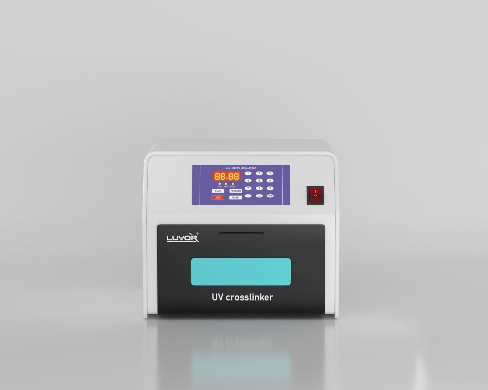
After transfer experiments such as Southern, Northern, Dot, Slot blot and EMSA, the nucleic acid has to be fixed to a solid support. The traditional method bakes the membrane in an 80 ℃ vacuum oven for about 2 hours; with 254 nm UV, the amino groups of a nylon membrane form stable covalent bonds with the thymine of DNA (or the uracil of RNA), and the whole process takes tens of seconds rather than hours, with a dried membrane exposed directly.
Wet a sheet of absorbent paper with transfer buffer (such as 10× SSC) and lay it flat on the floor of the exposure chamber.
Place the nylon membrane on the absorbent paper with the nucleic acid side facing up, so that UV reaches the nucleic acid directly.
On the control panel, press program to select energy mode, enter the fixing dose on the numeric keys and press enter to confirm.
Press start; the display shows the remaining energy and exposure stops automatically at the set value.
After the beep, remove the membrane and clean the chamber to remove residual salt.
Recommended parameters:
| Wavelength | Recommended dose | Exposure mode | Exposure time |
|---|---|---|---|
| 254 nm | 0.12 J/cm² (120 mJ/cm²) | Energy mode | 30-45 s |
Compared with traditional baking, UV crosslinking cuts the time substantially and usually improves the hybridization signal, which helps with low-abundance targets; and because energy mode stops on integrated dose, the result is not affected by the lower UV output of an aged lamp and is reproducible. Note that the UCL-3200 input unit is J/cm², so enter 0.12 directly for membrane fixing; other models may use mJ/cm² or μJ/cm²×100, and the conversion needs care.
Large DNA fragments in an agarose gel (typically above 20 kb) transfer poorly, so they need fragmentation before transfer to a nylon membrane. The traditional method uses HCl for depurination, but that reaction is hard to control and very temperature sensitive, and a slight error causes over-degradation.
Nicking the DNA in an ethidium bromide (EB) stained gel with 254 nm short-wave UV is more reliable and more repeatable, and it removes the steps of preparing and rinsing depurination reagents.
Recommended parameters:
| Wavelength | Mode | Reference dose | Sample |
|---|---|---|---|
| 254 nm | Energy mode | about 0.08 J/cm² (80 mJ/cm²) | DNA in EB-stained gel |
Aerosol contamination in PCR laboratories is a common cause of false positives. Exposing prepared reaction mixes or the associated consumables to 254 nm UV damages residual nucleic acid template and is a routine measure against false positives. The same instrument can also sterilize vessels, consumables and bench items by UV - one of the two core uses the UCL-3200 is specified for (eliminating PCR contamination and UV sterilization).
Arrange evenly: uncap or unwrap and lay items in a single layer in the exposure chamber so that every surface can receive UV.
Time mode: set about 0.5-1 minute (30-60 seconds); a beep signals the end and the unit returns to standby.
Suitable items: pipette tip boxes, centrifuge tubes, Petri dishes, small instruments and similar.
Note: UV travels in straight lines and does not penetrate, so it acts on directly exposed surfaces; shadowed faces and the inside of liquids need separate treatment. Where dose consistency matters, energy mode with periodic calibration is preferable.
Exposing cells or a cell suspension to a series of increasing UV doses builds a survival curve and a mutation frequency curve, a classical approach in mutation breeding and DNA damage research; place the sample at a moderate distance from the lamp so that the dose is even.
In microbial genetics, the RecA protein takes part in recombinational DNA repair, and a recA-deficient strain is markedly more sensitive to UV than the wild type. Applying the same UV dose to candidate strains and comparing survival allows screening and functional testing of recA mutations in E. coli.
The key to this kind of experiment is that every sample must receive the same cumulative dose, which is exactly where energy mode and real-time dose monitoring help: a programmable microprocessor monitors UV emission continuously and stops automatically at the preset energy, so runs on different batches and different days keep the same integrated dose and the data stay comparable.
UV exposure generates thymine dimers at defined sites in DNA, and these photoproducts block cleavage by restriction enzymes. Expose the DNA to UV to create dimers, then treat with restriction enzymes to obtain partial digestion products; the result can be used for rapid restriction map localization as a complement to classical gene mapping.
Tip: dimer yield is directly related to dose, so establish a low-dose gradient to find the working condition, and keep enzyme amount and reaction conditions identical across samples so that products can be compared.
In protein-nucleic acid interaction work, UV crosslinking can covalently fix the bound complex for later detection and verification, and can also be combined with workflows such as EMSA to stabilize the complex on a membrane for further analysis.
With protein samples, note that too high a UV dose can damage protein epitopes or reduce binding activity; start from a lower dose, optimize step by step, and include an unexposed control so that crosslinking efficiency and sample loss can be assessed.
UV is regarded as a supplementary disinfection method alongside chemical disinfectants in medical environments. With the energy mode of the UCL-3200, a cumulative dose of about 1 J/cm² (1000 mJ/cm²) can be set for supplementary disinfection - a level well above what routine surface disinfection requires, and suitable for instrument surfaces, small containers and items inside clean areas.
Even light: a mirror reflector and a matte reflector are used together to keep light intensity uniform across the whole chamber.
Real-time monitoring: a programmable microprocessor monitors UV intensity continuously, giving a repeatable dose regardless of place and time.
Traceable values: the built-in UV radiometer is traceable to CMA and CNAS laboratories, so dose records are straightforward to archive and review for quality purposes.
Boundary note: UV does not penetrate, so it acts on directly exposed surfaces; shadowed areas and the interior of porous structures need other disinfection methods, and on-site validation is advisable.
Beyond quantitative crosslinking, time mode supports process work that needs continuous exposure; the time range covers 00.00-99.59 minutes or 000.0-599.5 minutes, which spans controlled exposure from tens of seconds to several hours.
Gel polymerization: control of UV-induced gel polymerization.
Coating cure: UV curing and surface treatment of small parts.
Sample drying: assisted UV drying of thin-layer samples.
Materials testing: UV aging and weathering pre-assessment of small material samples.
Prohibited: never place flammable, explosive or volatile organic solvents in the exposure chamber for irradiation.
The table below summarizes the recommended settings for the eight scenarios as a quick reference for daily work. The values are general starting points; use the results of your pilot run. Note that the energy unit on the UCL-3200 panel is J/cm².
| Application | Wavelength | Recommended mode | Reference dose / time | Key point |
|---|---|---|---|---|
| Nucleic acid membrane fixing (Southern / Northern / Dot / Slot blot) | 254 nm | Energy mode | 0.12 J/cm² | Nucleic acid side up, facing the lamps; about 30-45 s |
| DNA nicking in agarose gel | 254 nm | Energy mode | about 0.08 J/cm² | Replaces HCl depurination before transfer of large fragments |
| PCR contamination control | 254 nm | Time mode | about 0.5-1 min | Damages residual template, lowers false-positive risk |
| Sterilization of vessels, consumables and bench surfaces | 254 nm | Time mode | about 0.5-1 min | Single layer, no shadowing; acts on exposed surfaces |
| UV-induced mutation | 254 nm | Time / energy mode | Gradient set by cell tolerance | Sample centered; cumulative dose must match across samples |
| recA function testing and mutant screening | 254 nm | Energy mode | Same dose, compare survival | Dose consistency is the precondition for comparable data |
| Thymine dimer gene mapping | 254 nm | Energy mode | Start with a low-dose gradient | Dimers block digestion; use partial products for mapping |
| Biomolecule crosslinking (protein-nucleic acid) | 254 nm | Energy mode | Start low, optimize by gradient | Avoid high doses that damage protein activity and epitopes |
| UVC surface disinfection | 254 nm | Energy mode | about 1 J/cm² | Real-time dose monitoring, even light, repeatable result |
| UV curing / polymerization / drying | 254 nm | Time mode | Set by process (up to 599.5 min) | Starts on the last program, one-key operation; no flammable solvents |
These scenarios cover steps from basic research to quality control. Typical users include universities and research institutes (molecular biology, microbiology and genetics laboratories), hospital clinical and pathology departments (membrane fixing and contamination control in molecular diagnostics), CDC and public health bodies (pathogen nucleic acid testing and laboratory contamination management), biologics and pharmaceutical QC (nucleic acid hybridization assays, small item handling in clean areas), third-party testing laboratories (standardized crosslinking of high-throughput samples) and food and agricultural research units (breeding mutagenesis, microbiological testing and UV treatment of materials).
UV causes irreversible damage to skin and the cornea, so strict protection is required when using a UV crosslinker:
Never open the door while the lamps are lit; confirm the door is fully closed before starting (most models have an interlock and will not start with the door unlatched).
Place the instrument on a stable, ventilated bench, away from humidity, high temperature and aggressive reagents.
Calibrate output intensity with a UV energy meter at intervals, and replace lamps when they reach rated life.
Clean the chamber after each use, remove residual salt and spills, and keep it dry.
Never place flammable, explosive or volatile organic solvents in the chamber for irradiation.
If a lamp fails to light, the interlock fails or the panel reports an error, switch off and stop using the unit, and contact service personnel; do not open the instrument yourself.
For detailed operating rules, follow the product manual supplied with the unit and your laboratory safety regulations.
From 0.12 J/cm² for nucleic acid membrane fixing to about 1 J/cm² for supplementary UVC disinfection, the value of a UV crosslinker is that it turns UV exposure into a metered, reproducible experimental step. With real-time microprocessor dose monitoring, a CMA/CNAS-traceable radiometer and a uniform mirror chamber, the UCL-3200 can move between these scenarios on one instrument; the manufacturer also offers free replacement for faults within one year and free repair within three years, which gives routine use more support.
To confirm the wavelength version for a specific scenario (UCL-3200M 302 nm / UCL-3200L 365 nm), dose settings, consumables and power requirements, contact our application engineers for selection advice.
This article belongs to the laboratory research techniques section.
Other sections on the same product line include UV transilluminators (gel and thin-layer chromatography excited from below, so bands can be read without turning the gel over), UV crosslinkers (dose set in J/cm², the figure that matters for both nucleic acid crosslinking and materials curing), and life science instruments (the entry point for fluorescence imaging, UV crosslinking, UV transmission and cell irradiation).
Models mentioned in this article: UCL-3200, LUV-450, XEPU-1126. Range, accuracy and accessory lists for each model are given on its product page.

Email: info@xinysun.com

Tel: 131-2093-6518

Address: 1525 Minqiang Road, Bldg 26, Songjiang, Shanghai

Copyright 2026 Xinysun Instrument (Shanghai) Co., Ltd. All rights reserved.
ICP:ICP-2026046447




 Industrial Test Instruments


