
Products
PRODUCTS Breadcrumb: Home > Products > Life Science Instruments > UV Transilluminators
 Overview
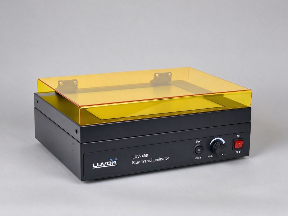
LUV-450 blue light transilluminator for SYBR Green and GelGreen gels: 470 nm LED excitation, white light mode, 10-99% intensity, 26x21 cm area, no UV.
The LUV-450 LED blue-light transilluminator, often called a blue-light gel cutter, uses 470 nm blue light instead of ultraviolet to excite fluorescent dyes, and 20 white LEDs switch it to white-light viewing. It addresses two long-standing problems of conventional UV transilluminators: the carcinogenic risk of ethidium bromide (EB) and the damage shortwave UV does to DNA samples.
Newer non-toxic dyes such as SYBR Green, SYBR Safe, and GelGreen have excitation peaks in the visible range, so a standard UV transilluminator cannot excite them; blue light is required. For laboratories that have already switched dyes, the LUV-450 is the unit that completes the move to safe dyes.
The traditional workflow uses EB staining and UV excitation, which brings the toxicity of EB and UV damage to samples. With safe dyes such as the SYBR series, both excitation and emission fall in the visible range, and the spectrum of a UV transilluminator no longer matches - the problem is not sensitivity, it is that the dye is not excited at all.
Excites SYBR Green, GelGreen, SYBR Safe, and similar dyes, and the bands fluoresce green. There is no ultraviolet output, so exposure risk to skin and eyes drops sharply and DNA damage stays low.
White LEDs (400-760 nm) support routine inspection: whether staining is even, whether the gel is intact, whether the wells are damaged. Confirming position in white light before cutting is the safer sequence.
One switch changes modes, so viewing, positioning, and cutting happen on the same instrument.
470 nm blue excitation means you do not need the precautions a UV transilluminator demands; goggles plus standard protection are enough.
Visible light carries less energy than UV, so DNA damage is low and recovered fragments clone and ligate more reliably.
Blue light excites the bands, white light shows the gel condition. Flip the switch; no second instrument or lamp change is needed.
Set it to match dye concentration and band brightness: dim bands brighten, strong bands avoid overexposure.
Imported LED emitters last long and need no maintenance, which removes the consumable cost of periodic UV tube replacement.
Illumination is even across a 26×21 cm area, so photographs show no dark corners and several small gels side by side read the same.
Selection guidance: start with the excitation wavelength in the dye datasheet. A peak near 460-480 nm works in blue mode; if the peak sits further toward the red end, assess signal-to-noise before you decide.
The LUV-60 amber glasses ship with the unit and cut the blue background to raise band contrast. Put them on before you switch on blue light.
Switch to white mode and confirm the gel is intact, the wells are undamaged, and roughly where the bands sit. Then switch to blue for exact positioning.
Start low and increase until the bands read clearly. Prolonged high-intensity blue light also bleaches fluorescence, and faint bands are affected before strong ones.
Blue light damages DNA less, but recover and purify the slice soon after cutting rather than leaving it on the illuminated surface.
Wipe the surface with a soft lint-free cloth and avoid scratching the filter plate; dried residue degrades transmission uniformity.
It depends on the dye. For dyes whose excitation peak sits in the visible range, such as the SYBR series, blue excitation is the better match and sensitivity is comparable to UV. For dyes that need UV excitation, such as EB, blue light excites poorly and does not apply. The reference point is the excitation wavelength in the dye datasheet, not a general impression that blue is weaker than UV.
Not recommended. The LUV-450 is designed to move away from EB and UV: its value appears once you switch to safe dyes such as SYBR Safe and GelGreen. If EB still dominates your workflow, choose a UV transilluminator, for example the LUV-260 302 nm model.
It is not redundant. Confirming gel integrity, well condition, and approximate band position in white light before cutting shortens the time spent hunting under blue light. For imaging records you can also capture a white-light overview, then a blue-light fluorescence image. A white-light image is useful supporting material for gel documentation.
No fixed replacement interval applies. LEDs last long, degrade slowly in normal use, and do not blacken at the ends the way tubes do. If band brightness falls noticeably under the same conditions, check filter plate cleanliness and dye condition before you suspect the light source.
Yes. The two are close in size (325×322×105 mm), but keeping both is not necessary: once the dye switch is complete, UV transilluminator use drops sharply. Most laboratories keep one UV unit for legacy EB workflows or special needs and run everyday viewing and cutting on the LUV-450.
Compatibility table for the LUV-450 and common safe dyes
Guidance for converting a workflow from EB plus UV to SYBR plus blue light
Filter and exposure settings for gel photography under blue light
Blue-light gel cutting procedure and protective equipment checklist
LUV-60 amber viewing glasses ship with the unit, so it is ready to use on arrival
Advice on dye matching and workflow conversion
Long-term supply of filter plates and other parts
Demonstration units and operator training available
Send inquiry

Email: info@xinysun.com

Tel: 131-2093-6518

Address: 1525 Minqiang Road, Bldg 26, Songjiang, Shanghai

Copyright 2026 Xinysun Instrument (Shanghai) Co., Ltd. All rights reserved.
ICP:ICP-2026046447




 Products
 Applications
 Previous:
 Back to list

 Industrial Test Instruments


