
Products
PRODUCTS Breadcrumb: Home > Products > Life Science Instruments > UV Transilluminators
UV transilluminator selection: benchtop models cover 254/302/365 nm with single or dual wavelength for nucleic acid gel observation and gel excision and recovery; UV transillumination tables use special UV filter glass for even, ng-level sensitivity; gel transilluminators have adjustable intensity for imaging; use a 470 nm blue LED cutter with safe dyes such as SYBR. Models, parameters and quotations from Xinysun.
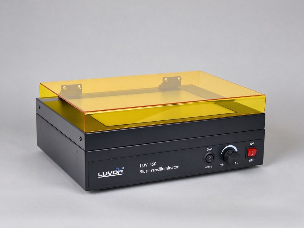
 Model: LUV-450

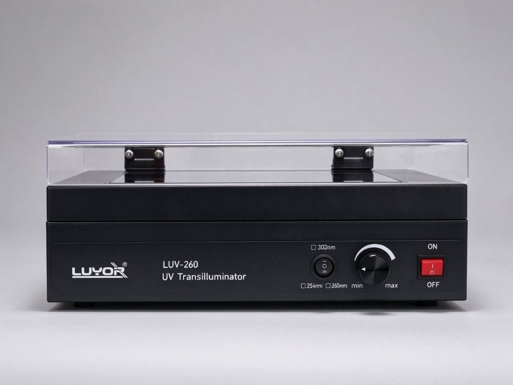
Model: LUV-260
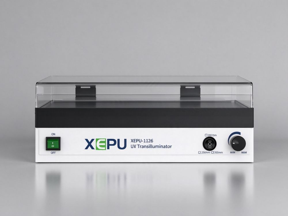
Model: XEPU-1126
Choosing a transilluminator comes down to two things: which wavelength to use, and what gel size the surface can accommodate. Use 302nm (UV-B) for viewing bands - dyes such as EB and GelRed are excited efficiently and give bright bands. Use 365nm (UV-A) for gel excision and recovery: the energy is lower, DNA damage is reduced and recovered fragments stay more intact. Dual-wavelength models switch between viewing and excision without repeatedly moving the gel, and labs already using non-toxic dyes can simply use a blue-light transilluminator instead of UV.
| Model | Wavelength | Illumination Area | Intensity Control | Notes |
|---|---|---|---|---|
| LUV-260 | 254 / 302 / 365nm, single or dual wavelength optional, 8×8W tubes | 26×21cm | - | ng-level sensitivity, adjustable safety shield, fan cooling |
| XEPU-1126ML | 4×302nm + 4×365nm (8W each) | 26×21cm | 5%-99% | One unit handles both viewing and excision; hinged safety shield is adjustable |
| LUV-450 | 24×470nm blue LEDs + 20 white LEDs | 26×21cm | 10%-99%, switchable blue/white | Compatible with non-toxic dyes such as SYBR Green; no UV, no EB |
The comparisons above are compiled from the published specifications on each product page. For exact configurations, prices and lead times, please refer to the relevant product page or call the number at the bottom of the page. Xinysun Instruments (Shanghai) Co., Ltd. supplies LUYOR and XEPU instruments for industrial inspection, UV curing and speed measurement.
A transilluminator mounts the UV source below the surface, with the gel placed directly on the illuminated plate. UV light passes through the gel and excites the DNA dye to fluoresce, making the bands visible. Its key specifications are the uniformity of illumination across the surface and ease of operation - the smaller the brightness difference between the edge and center of a gel, the more reliable size determination and quantification become.
It is a different type of instrument from a UV crosslinker: a transilluminator has an open surface for viewing, gel excision and photography, while a crosslinker has a closed chamber for delivering a measured dose for membrane crosslinking. Substituting a transilluminator's 'irradiate for a while' for crosslinking gives an uncontrolled dose and poor reproducibility.
1) Pre-run: switch on the lamp and let it stabilize for a few minutes until the intensity plateaus, then load the gel.
2) Load the gel: place it face up and flat against the surface, removing air bubbles and excess buffer, since bubbles can reflect light and appear as false bands.
3) Adjust the intensity: start low and increase gradually to find a setting where bands are clear without background overexposure; lowering the intensity during excision reduces the time DNA is damaged.
4) Use the safety shield: pull the shield into the viewing position and observe through the window; wear a face mask and gloves when excising and work as quickly as possible.
5) Document with a photo: fix the aperture and exposure, add a filter matched to the wavelength, and use the same settings for all gels in a batch so brightness can be compared later.
Once these three things are settled, the model is essentially determined.
| Requirement | Recommended Configuration | Reason |
|---|---|---|
| Viewing bands and determining size | 302nm, adjustable intensity | EB and GelRed are excited efficiently, giving bright bands |
| Gel excision and recovery | 365nm or switchable dual wavelength | Lower energy, less DNA damage, intact recovered fragments |
| Both viewing and excision | 302+365nm dual-wavelength model | No repeated moving of gels, reducing cross-contamination |
| Non-toxic dye workflows | 470nm blue-light transilluminator | No UV exposure risk; compatible with dyes such as SYBR Green |
| Surface size | Allow margin for the largest gel; 26×21cm is common | If the gel extends beyond the illuminated area, edge bands will appear dim |
- Agarose gel electrophoresis: viewing bands of PCR products, restriction fragments and plasmids.
- Gel excision and recovery: locating the target band at 365nm to reduce UV damage time.
- Protein and Native PAGE: observing bands and migration with appropriate dyes.
- TLC plates: observing fluorescence-quenching spots at 254nm.
- Teaching labs: multiple observers at once; the safety shield and low-intensity setting are safer.
- Photography and archiving: recording gel images with fixed settings for later quantification and review.
- Selection and brands: How to Choose a UV Crosslinker and Fluorescent Protein Excitation Source? A Guide to UV and Fluorescence Tools for Life Sciences, Blue-Light vs. UV Transilluminator: Dye Compatibility and Protection for 470nm Gel Excision, How to Choose a UV Transilluminator? 254/302/365nm Wavelengths, Tube Count and Excision...
- Operation and interpretation: Vanillin Rapid-Test Strips: The 365nm of the LELMS-38L Tri-Wavelength UV Lamp...
- Field and literature case studies: Eight Applications of the UCL-3200 UV Crosslinker
Use 302nm (UV-B) for viewing bands: fluorescent dyes are excited efficiently and bands are bright, suiting size determination and quantification. Use 365nm (UV-A) for gel excision and recovery: the energy is lower, DNA damage is reduced and recovered fragments stay more intact. Dual-wavelength models switch between the two, avoiding cross-contamination from repeatedly moving the gel.
For safety and dye compatibility it can replace UV in some scenarios. Blue light at 470nm works with non-toxic dyes such as SYBR Green, GelGreen and SYPRO Ruby, with no UV exposure risk and no DNA damage, suiting frequent use and teaching demonstrations. However, legacy protocols still using EB, or experiments that need to excite UV-specific bands, still require a UV transilluminator.
A transilluminator's UV light shines upward toward the operator's face and eyes, so a safety shield is basic safety equipment. Pull the shield into the viewing position and observe through the window; when excising, wear a face mask and gloves and keep exposure as short as possible. A hinged, height-adjustable shield is more convenient than a fixed one for working with gels of different thicknesses.
Increase the intensity rather than the exposure time first, then consider switching to 302nm (more efficient excitation than 365nm). Thick gels absorb significantly, so you can lower the gel concentration, make the gel thinner, or extend staining and destaining times. If the edges look dark and the center bright, surface uniformity is insufficient; move the gel toward the center and reduce the observation area.
After each use, first aspirate the buffer, then wipe with 70% ethanol or a detergent, and finally wipe clean with pure water. If there is a risk of amplified-product residue, run the UV with no gel present for 10-15 minutes to decontaminate the surface. Avoid wiping the quartz surface with chlorine-based disinfectants over the long term, as this can cause hazing that reduces light transmission.
Semi-quantification is possible, but it requires gels from the same batch, the same exposure and the same dye concentration, plus a Marker of known amount as an internal reference; saturated pixels and overexposed areas cannot be quantified. For rigorous quantification use a cooled imaging system with dedicated software; phone photos are suitable only for documentation and within-group comparison.
It depends on the dyes and existing protocols. Dyes such as SYBR Green, GelGreen and SYPRO Ruby are excited well by 470nm blue light, do not damage DNA and require no UV protection, suiting routine and teaching use. However, legacy protocols still using EB, or observing TLC plates by fluorescence quenching at 254nm, still require a UV transilluminator.

Email: info@xinysun.com

Tel: 131-2093-6518

Address: 1525 Minqiang Road, Bldg 26, Songjiang, Shanghai

Copyright 2026 Xinysun Instrument (Shanghai) Co., Ltd. All rights reserved.
ICP:ICP-2026046447




 Products
 Applications
 Industrial Test Instruments


