
Applications
 Breadcrumb: Home > Applications > Lab Research
 Updated:2026-11-02
 Views:
In August 2026, a team at Guangxi University published a macadamia (Macadamia spp.) transformation study in Plants (15(16), 2418): Agrobacterium rhizogenes K599 delivered in planta hairy-root transformation, combined with four visual reporter genes (DsRed2, eGFP, RUBY, AtPAP2), without antibiotics and without tissue culture. The work reports positive-root rates of 39.25%-47.38% with injection on seedlings at OD₆₀₀ = 0.5, and 24.8%-28.2% on mature trees at OD₆₀₀ = 1.0. This article covers where the advance lies, which step the LUYOR-3415RG handles, how to choose among the four reporters, a set of transformation parameters that can be followed directly, and the roughly 30-day timeline.
In August 2026, the Xu Zengfu team at the College of Forestry, Guangxi University, published a study on genetic transformation of macadamia (Macadamia spp.) in MDPI Plants (Plants 2026, 15(16), 2418, open access). Macadamia is an important economic nut crop, but transformation has long been difficult to carry out, which holds back functional genomics and molecular breeding. The paper uses Agrobacterium rhizogenes K599 for in planta hairy-root transformation, combined with four visual reporter genes, to screen positive roots without antibiotics and without destroying the material - and the eGFP and DsRed2 fluorescence observations in the paper were made with the LUYOR-3415RG dual-wavelength fluorescent protein excitation source.
For labs working on woody plant transformation, the paper carries two kinds of value: it turns an in planta route into reproducible parameters, and it shows how a three-layer screen - pigment marker, fluorescent reporter, molecular confirmation - is chained together. This article takes those two threads apart and explains which step the 3415RG covers.
Conventional plant transformation starts with in vitro culture: explant sterilization, callus induction, shoot regeneration, each step genotype-sensitive, and woody species are especially hard. The paper takes the in planta route instead, letting A. rhizogenes induce hairy roots directly at wounds on the hypocotyl of an intact plant and removing the sterile-culture step. Three key advances:
Reporters made visible. Four systems run in parallel: DsRed2, eGFP, RUBY and AtPAP2. DsRed2 and eGFP give red and green fluorescence, RUBY gives betalain, and AtPAP2 acts through the anthocyanin pathway; the latter two are visible to the naked eye, the former two are read with an excitation source.
Pushed out to mature field trees. Air layering was introduced, using the girdled wound as the infection site, and transformation was completed on branches of 5-year-old grafted trees. Comparable systems had largely stayed at the seedling stage, and tissue maturity has long been a barrier that woody transformation cannot avoid.
Not genotype-picky. Seedlings came from open pollination, so the genetic background is highly diverse (four cultivars: A4, GR1, HAES900, O.C.). The rol gene cluster amplifies cell sensitivity to hormones and narrows the efficiency gap between genotypes; positive hairy roots were recovered from all four cultivars.
Four reporter genes map onto four read-outs, and the division of labor in the paper is clear:
| Reporter gene | Read-out | Equipment used | Observation in the paper |
|---|---|---|---|
| eGFP | Blue excitation, green fluorescence | LUYOR-3415RG (R band 440-460nm) | Excitation/emission peaks about 488/507nm, used in cv. O.C. |
| DsRed2 | Green excitation, red fluorescence | LUYOR-3415RG (G band 510-540nm) | Excitation/emission peaks about 558/583nm, used in cv. GR1 |
| RUBY | Red seen directly by eye | Ambient white light or bright-field stereo microscope | Positive roots dark red or purple-red; non-transgenic roots milky white or light brown |
| AtPAP2 | Purple seen directly by eye | Ambient white light or bright-field stereo microscope | Positive roots pale purple or light red, dependent on the endogenous anthocyanin pathway |
| High-resolution confirmation | Fluorescence stereo microscope | Nikon P-DSL32 | Image capture and read-out down to subcellular localization |
The table implies a practical workflow: scan the living material quickly with the 3415RG, pick out the suspicious positive roots, then send them to the microscope for high-resolution confirmation. The paper does not put light intensity, exposure time or working distance into the methods - this is a viewing and imaging tool, not a quantitative irradiance instrument, and the parameters just need to satisfy "can it be seen". What does belong in the methods is the excitation band and the filter configuration.
The discussion compares the four systems point by point, and that comparison is useful when picking a marker:
| System | Strengths | Limitations |
|---|---|---|
| eGFP / DsRed2 | Supports subcellular localization and high-resolution histology, and avoids interference from endogenous pigments | Depends on fluorescence equipment, awkward in the field and in high-throughput work; signal fades in aging roots |
| RUBY (betalain) | Uses an endogenous tyrosine-derived pigment, so it is unaffected by the recipient's secondary-metabolism background, and it is visible to the naked eye | Pigment degrades after prolonged culture; stress-induced anthocyanins can interfere |
| AtPAP2 (anthocyanin) | Macroscopically visible, needs no equipment at all | Depends on coordinated expression of the recipient's endogenous anthocyanin pathway; the specific expression pattern in macadamia roots limits color development, and the signal is weaker than RUBY |
The paper's recommendation is to combine all three layers: pigment markers for coarse field screening, fluorescent reporters for lab confirmation and imaging, and RT-qPCR plus genomic PCR for the molecular verdict. Any one layer alone is not enough - pigment degrades, fluorescence fades, and molecular evidence is the hard part.
The methods section is fairly detailed; the parameters below can be copied straight into a lab notebook:
| Step | Parameters in the paper | Notes |
|---|---|---|
| Strain | A. rhizogenes K599 | Carrying pKSE402-eGFP, p501-SUC2::JcFT-DsRed2, p35S-RUBY, pRUBY-WIP, pGNP-AtPAP2 and other constructs |
| Bacterial preparation | Centrifuge at 8000×g for 2 min, resuspend | Grown on YEB solid medium at 28°C for 36-48 h to form a lawn |
| Infiltration buffer | 10 mM MES + 10 mM MgSO₄ + 100 μM acetosyringone | pH 5.5-5.6, pre-induced standing in the dark at 28°C for 1 h |
| Bacterial density (seedlings) | OD₆₀₀ = 0.5 | For air layers on mature trees, OD₆₀₀ = 1.0 works better; see the next section |
| Inoculation | Injection (INJ) or dip (DIP) | Hypocotyl cut about 1 cm below the cotyledonary node, wound facing up, replanted in vermiculite |
| Co-cultivation | 28°C, dark, 3 days | Return to a normal photoperiod after co-cultivation |
| Culture conditions | 26°C, 16 h light / 8 h dark | Misted daily to hold humidity |
| Air layering | Girdle about 1 cm wide down to the cambium, cotton wool soaked in bacterial suspension | 5-year-old grafted trees, shoot diameter 1.0-1.5 cm, wrapped with a rooting box and sterile substrate |
| Assessment time points | 25 days after seedling inoculation; 30 days after air-layering transformation | Open the box to check rooting and fluorescence signal |
| Molecular confirmation | RT-qPCR (reference gene Actin1, 2-ΔΔCt method) + genomic PCR | n = 5 positive roots taken at random, 1.5% agarose gel electrophoresis |
The paper ran two key comparisons, and the conclusions can guide practice directly. Start with inoculation method (Figure 2B):
| Inoculation method | Positive induction rate | Difference |
|---|---|---|
| Dip (DIP) | 27.93%-30.84% | Baseline |
| Injection (INJ) | 39.25%-47.38% | Significantly higher than DIP in GR1 and O.C. (p < 0.01) |
The authors' explanation: a needle wound is mechanically deeper, so the suspension reaches the vascular cambium and the deeper parenchyma cells directly, and those are the tissues where hairy roots initiate most readily. Dipping acts on the cut surface alone.
Second, bacterial density for air layering on mature trees (Figure 5):
| OD₆₀₀ | GR1 | JW | O.C. | Reading |
|---|---|---|---|---|
| 0.5 | 17.7% | 17.7% | 15.7% | Bacterial load too low |
| 1.0 | 26.0% | 28.2% | 24.8% | Balance between efficiency and tissue vigor |
| 1.5 | 21.6% | 21.0% | 15.3% | Too dense, biological stress, efficiency falls back |
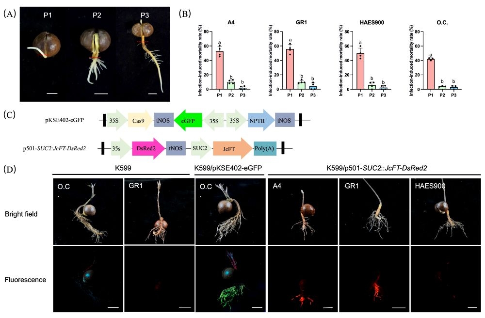
One more easy trap: seedling developmental stage. The paper divides the hypocotyl into P1 (just breaking the seed coat), P2 (hypocotyl elongating, lateral roots appearing) and P3 (clearly lignified). Infection at P1 carries 42%-56% lethality; at P2 it drops to 4%-10%. Tissue that is too young cannot take the infection, tissue that is too old has lignified, and P2 is the useful window. GR1 also shows a clear developmental dependence - its positive rate at P1 is significantly higher than at P3.
One control datum worth noting: air layers treated with sterile buffer alone (mock) produced 3.3, 2.3 and 2.7 roots per shoot in the three cultivars; with commercial rooting powder every shoot produced 8 roots, yet the transformation rate was 0. More roots does not mean successful transformation, which is exactly why reporter read-out is needed.
The paper recorded morphological changes 17, 30, 40 and 65 days after air layering; that timeline can be used directly to schedule observation points:
Day 17: tissue above the girdle swells and primary callus begins to form, with no roots visible yet;
Day 30: abundant dense callus and clear adventitious roots - this is a suitable window for initial fluorescence screening, since roots have emerged and the signal is not yet masked by aging tissue;
Day 40: roots enter rapid elongation, the root system thickens, and single-root signal is still clear;
Day 65: root number rises further and a fairly complete root system forms, so several root segments can be examined at multiple points.
The paper notes that positive roots appear on average about 30 days after inoculation. Under the fluorescence microscope, clear red fluorescence is visible inside the roots, with DsRed2 expressed mainly in vascular tissue; RT-qPCR shows JcFT and DsRed2 transcripts in transformed GR1, JW and O.C. all significantly higher than in controls (p < 0.01).
One negative result deserves attention: despite high JcFT expression, no apical flower-bud differentiation was observed within two months of inoculation. The authors suggest that FT protein is diluted by the large biomass before it reaches the shoot apex, and that mature tissue would need epigenetic remodeling before it can respond to a floral signal - the paper states clearly that this is a hypothesis, not yet verified.
Combining the paper's practice with the design of the instrument gives three concrete actions. Start with the instrument: the LUYOR-3415RG puts two excitation bands into one handheld unit, with two switches on the lamp body. In the standard configuration the R band is royal blue 440-460nm with a 500nm filter, which matches eGFP at 488/507nm; the G band is green 510-540nm with a 600nm filter, which matches DsRed2 at 558/583nm - which is why the paper used blue light for O.C. and green light for GR1. The two bands total 12 × 3 W LEDs, 27 W overall, an illuminated diameter of 180mm at 150mm from the lamp, net weight 0.9kg.
No need to open the culture vessel. Hairy roots are often kept in the vessel after induction, and opening it to look carries contamination risk. The 3415RG is a handheld external source: shine it through the vessel wall and fluorescence on the roots is visible without disturbing the material. For woody species whose transformation efficiency is already low this matters - one contamination event can ruin a batch.
In situ observation of roots. Macadamia hairy roots grow in vermiculite or substrate, and pulling them out damages them. Move the plant with its substrate into the dark, hold the source against the substrate surface, and positive roots show green or red fluorescence against a dark background. In the paper DsRed2 is expressed in vascular tissue, so the signal is concentrated and well bounded, which suits this non-destructive read-out.
Coarse screening on living field trees. Air layering is done in the field, and shoots cannot be carried back to the lab when the box is opened. The 3415RG weighs 0.9kg and runs from a rechargeable battery pack (AC 100-260V charging), so it can be taken to the tree. The mature-tree trials ran in Nanning from September to November, mean temperature 24-25°C, relative humidity 72%-78%, and in that setting portability counts for more than resolution.
Why not use a fluorescence microscope at this step? The paper itself hints at the answer: the field of view is small, the root system is an irregular three-dimensional structure, and scanning point by point is expensive in time; screening asks just one thing - "is this root positive" - and that needs no subcellular resolution. One user comment collected on the product page, from Huazhong Agricultural University, reads: "Roots of transiently transformed plants photographed with the 3415 also look good; the ability of this product to see red fluorescence exceeds that of the lab fluorescence microscope." Red fluorescent proteins are often limited by filter-cube configuration under a microscope, and a high-power dedicated source leaves more room. That said, images for publication are still better taken with a microscope or re-shot with a photographic filter - the two are complementary.
Settle the reporter gene, then the excitation band. eGFP means the R band, DsRed2/mCherry means the G band, and both means the standard 3415RG. Note that the two bands of the 3415 are fixed at the factory and set at order and cannot be changed on site - list every fluorescent protein the project involves before choosing.
Write the three-layer screen into the workflow. Pigment markers (RUBY/AtPAP2) for coarse field screening, the 3415RG for fluorescence confirmation on living tissue, RT-qPCR and genomic PCR for the molecular verdict. A line counts as positive after all three.
Watch for the P2 stage. Schedule seedling infection when the radicle and hypocotyl are elongating and lateral roots are just appearing, avoiding the 42%-56% lethality of P1 and the lignification of P3.
Injection beats dipping. Where conditions allow, use a 1 mL syringe to inject the suspension into the hypocotyl cut face from several directions; the positive rate rises from 27.93%-30.84% to 39.25%-47.38%.
Match bacterial density to the situation. OD₆₀₀ = 0.5 for seedlings, OD₆₀₀ = 1.0 for air layers on mature trees; do not simply go denser, since at 1.5 efficiency falls back to 15.3%-21.6%.
Put observation points on the calendar. Check seedlings 25 days after inoculation, run initial fluorescence screening on air layers at day 30, and revisit root segments at days 40 and 65. Get into the habit of recording band and filter with each observation so the work can be traced.
The division of labor is different. The microscope has a small field of view and the root system is an irregular three-dimensional structure, so point-by-point scanning is expensive in time, while screening needs just a yes/no call on positivity. A handheld excitation source can shine through a vessel, through substrate, or in the field, without damaging the material and quickly. High-resolution imaging and subcellular localization still belong to the fluorescence stereo microscope, such as the Nikon P-DSL32 used in the paper.
The R band is royal blue 440-460nm with a 500nm filter, for GFP/eGFP/fluorescein, matching eGFP at 488/507nm; the G band is green 510-540nm with a 600nm filter, for DsRed/TdTomato/mCherry, matching DsRed2 at 558/583nm. Both switches sit on the lamp body, so changing band needs no lamp swap and no realignment.
Injection on seedlings reaches 39.25%-47.38%, while air layering on mature trees at OD₆₀₀ = 1.0 gives 24.8%-28.2% (JW at the high end, 28.2%). The gap comes from tissue maturity: mature shoots are more lignified and divide less actively, and girdling can rebuild a hormone-sensitive microenvironment in part. The value of the mature-tree system is that it bypasses years of nursery propagation, so a lower efficiency still pays.
No. The paper's gradient test shows the positive rate rising clearly as OD₆₀₀ goes from 0.5 to 1.0 (GR1 from 17.7% to 26.0%), then falling back at 1.5 (GR1 21.6%, O.C. 15.3%). Too dense a suspension brings biological stress, tissue vigor drops, and the transformation window narrows.
Each suits a different situation, and both are needed. RUBY works through betalain pigmentation, needs no equipment, and suits field work and large-scale coarse screening; but the pigment degrades after prolonged culture, and stress-induced anthocyanins can interfere with read-out. The paper recommends combining the three layers - pigment marker, fluorescent reporter, molecular confirmation - with the 3415RG or a microscope at the fluorescence layer and RT-qPCR plus genomic PCR at the molecular layer.
LUYOR-3415RG dual-wavelength fluorescent protein excitation source - product page: R 440-460nm for GFP, G 510-540nm for DsRed, 12 × 3 W LEDs, net weight 0.9kg;
PsLykX gene-editing verification and hairy-root fluorescence screening in pea - the same instrument in a legume hairy-root system;
Culture-free hairy-root transformation in raspberry: how to screen eGFP-positive roots quickly - fluorescence read-out details for culture-free routes;
Positive screening of soybean hairy roots - the screening workflow in a GmSIN1 salt-tolerance study;
Positive screening of Salvia miltiorrhiza hairy roots - DsRed labeling in medicinal-plant synthetic biology;
eGFP colony fluorescence counting in a 2025 Nature alfalfa study - fluorescence counting at colony level;
LUYOR-3415 dual-wavelength scheme: high-throughput initial screening of living transgenic material - the five-band code system and combination selection;
Six fluorescence observation scenarios for the 3415RG - practical use in culture vessels, the field and seed sorting.
Reference: Mo Y, Fei YC, Tian X, et al. Efficient In Planta Induction of Transgenic Hairy Roots in Macadamia Seedlings and Mature Trees Using Visual Reporters. Plants 2026, 15(16), 2418. DOI: 10.3390/plants15162418 (CC BY open access). All data cited here come from that paper; for specific experimental conditions refer to the original paper and the instrument manual.
This article belongs to the research laboratory techniques section.
Other sections on the same product line include GFP fluorescence flashlights (models sorted by GFP/RFP/DsRed band, handheld and benchtop) and life science instruments (the entry point for fluorescence imaging, UV crosslinking, UV transmission and cell irradiation).
Models mentioned in this article: LUYOR-3415RG, LUYOR-3421, LUYOR-3260RB. Ranges, accuracy and accessory lists for each model are given on the product pages.

Email: info@xinysun.com

Tel: 131-2093-6518

Address: 1525 Minqiang Road, Bldg 26, Songjiang, Shanghai

Copyright 2026 Xinysun Instrument (Shanghai) Co., Ltd. All rights reserved.
ICP:ICP-2026046447




 Industrial Test Instruments


