
Applications
 Breadcrumb: Home > Applications > Lab Research
 Updated:2026-11-02
 Views:
Genetic transformation in Rubus has long been limited by two barriers: strong genotype dependence and the need to pass regeneration through tissue culture. A protocol published in Horticultural Plant Journal in 2026 uses Agrobacterium rhizogenes strain K599 to bypass tissue culture, obtaining transgenic plants in three species and plantlets in 4-6 months under non-sterile conditions. The article focuses on the eGFP prescreen: just 61.36% of hairy roots scored as positive were confirmed by PCR, so 38.64% were false positives. It also sets out the system's key parameters, the explant preferences of each species, the operation of the fluorescence screen under blue light, and five common sources of false positives.
Genetic transformation in Rubus has long been stuck at two points: strong genotype dependence, and regeneration that has to go through tissue culture. In September 2026, Horticultural Plant Journal published online a protocol that uses Agrobacterium rhizogenes K599 to bypass tissue culture, obtaining transgenic plants in wild Rubus niveus, R. ellipticus and cultivated blackberry R. fruticosus, with plantlets formed in 4-6 months under non-sterile conditions. One data set is worth pulling out on its own: of hairy roots scored positive for eGFP, just 61.36% could be confirmed by PCR, and the other 38.64% were false positives. This article sets out the key parameters of the system and unpacks the practical points of the eGFP prescreen step.
Rubus contains more than 700 species, but just a few - raspberry, blackberry and a handful of others - are grown commercially. Wild germplasm goes unused not because it lacks value but because it cannot be transformed: the conventional Agrobacterium-mediated route demands strict sterility, a stable explant regeneration system and a selection cycle that often runs a full year, and many wild genotypes stall at the starting stage.
This protocol takes a different entry point. The T-DNA of the Ri plasmid in Agrobacterium rhizogenes carries three genes, rolA, rolB and rolC, and infection produces large numbers of hairy roots from the wound site. Two features of hairy roots are particularly useful for transformation: they develop from single cells, so the roots that emerge are not chimeric; and they grow without exogenous hormones, which means no carefully formulated medium and no strictly sterile environment. Rubus itself suckers from its roots, and hairy roots cultured for a period in a moist substrate can induce adventitious buds and so give whole plants.
Compared with the conventional route, what is removed is exactly the most labor-intensive stretch: no culture vessels, no repeated subculture in a laminar flow hood, no antibiotic selection marker. The cost is that the cycle is still not short - plantlets appear in 4-6 months under non-sterile conditions, about half of the roughly one year needed by traditional methods.
The study compared four variables systematically - strain, bacterial concentration, number of axillary buds and infection time - and the conclusions are fairly clear:
| Parameter | Range compared | Result |
|---|---|---|
| Strain | K599, C58C1, R1000 | K599 gave 56.5% induction, above 38.1% for C58C1 and 29.2% for R1000 |
| Bacterial concentration | OD600 0.2-1.0 | At OD600 = 0.6 induction reached 65.8%, the peak of the range |
| Number of axillary buds | Single, double, triple | Single-bud segments 79.2%, double 65.8%, triple 53.6% |
| Infection time | 5-20 min | No significant difference within the range; 5-10 min is enough |
| Cocultivation substrate | Moist vermiculite | Non-sterile conditions, no hormones or antibiotics needed |
That single buds outperform multiple buds runs against intuition but makes sense: more buds means a longer stem segment, a greater distance from wound to vascular bundle and less uniform penetration of the bacterial suspension; a single-bud segment is short, the infection surface is concentrated and rooting is more even. In practice, discard both the young tip of the shoot and the heavily lignified base, and take the middle portion of moderate maturity.
The suitable explant is not the same across the three species, which is an easy place to go wrong when designing experiments:
| Species | Suitable explant | Hairy root induction rate | Remarks |
|---|---|---|---|
| R. fruticosus (cultivated blackberry) | Stem segment | 81.8% | Root segments also work, about 33.33% |
| R. niveus (wild) | Stem segment | 80.5% | Petioles 11.58% |
| R. ellipticus (wild) | Petiole | 22.33% | Stem segments failed, browning rapidly after inoculation |
The reason R. ellipticus stem segments failed is stated directly: high polyphenol content and strong polyphenol oxidase activity cause the wound to oxidize rapidly, and the tissue browns and dies before the bacteria get in. Its petioles oxidize more slowly, which leaves a 22.33% chance. For genotypes like this, the direction for further gains is antioxidant pretreatment, but the paper notes that this step remains to be validated.
Another comparison concerns the infection method. In vitro stem segment infection reached 80.5%, while field air layering reached 41.9%. The difference lies in whether the wound really reaches the vascular region: the cross cut in layering often stops at the cortex, and outdoor temperature and humidity are uncontrolled, so callus formation is unstable. So even if the aim is to stay out of the laboratory, start with in vitro stem segments.
The choice of reporter gene is the other main thread of the paper. RUBY is a tandem of three betalain biosynthesis genes (CYP76AD1, DODA and a glycosyltransferase) that use tyrosine as substrate; positive tissue turns red directly and can be scored by eye without any excitation light. eGFP, by contrast, needs external excitation.
The two reporters perform quite differently:
| Indicator | RUBY | eGFP |
|---|---|---|
| Scoring method | Red color by eye | Green fluorescence under excitation light |
| Agreement between visual positive and PCR | Full agreement, no false positives found | 61.36% confirmed by PCR, 38.64% false positives |
| Fading over time | Red weakens or disappears in regenerated plants, but PCR stays positive | Fluorescence intensity fluctuates with expression level |
| Equipment needed | None | Excitation light source or fluorescence microscope |
RUBY's advantage is obvious at the early screening stage, but its fading also deserves attention. The explanation offered in the discussion is that Rubus is not in the Caryophyllales and lacks a cellular environment for stable betalain accumulation; several polyphenol oxidases and peroxidases promote pigment degradation; and betalain accumulation correlates positively with auxin level, so as auxin falls in mature tissue the red fades. Fading red therefore does not mean transformation failed, and conversely red cannot serve as the sole criterion - PCR is still needed.
The problem on the eGFP side is more direct: seeing green light does not mean a true positive. A 61.36% agreement rate means one in every three green roots is false. That proportion is high enough that, without confirmation, downstream gene function conclusions will be contaminated.
In the paper, eGFP was scored visually - looking at whether roots turn green under excitation light. The step looks simple, but in practice inconsistent scoring criteria are one of the main reasons for the high false positive rate. Following the usual instrument-side practice, it can be split into five steps:
The excitation maximum of eGFP is at 488 nm, so use the blue band (440-460 nm) rather than the ultraviolet band. A 365 nm UV lamp will also make GFP emit faint fluorescence, but eGFP is a red-shifted engineered protein and UV excitation is inefficient; the signal is weak enough that you have to dim the room and observe close up, which easily turns background noise into a positive call.
Starting 2-4 weeks after infection, when hairy roots have grown to 2-3 cm, is a reasonable time to begin scoring. Roots that are too short break easily during handling; roots that are too old have suberized epidermis that blocks fluorescence. Hairy roots of R. niveus become slightly lignified after about 3 months in culture and are thinner than wild-type roots, so scoring at this stage needs more care.
A high-power LED source can excite distinguishable fluorescence under ordinary room lighting; move the culture box away from the window and switch off direct overhead lamps. Complete darkness is actually harder to work in - taking roots and labeling them both become error-prone.
Include untransformed wild-type roots from the same batch as a negative control, confirm that negative roots show no green from any angle, and begin scoring samples after that check. This step rules out three kinds of interference: autofluorescence of the substrate, epiphytic algae and filter crosstalk.
For each root scored positive, note on the spot which plant it came from, its orientation and approximate length, then cut a 1-2 cm segment for PCR. The paper used a 559 bp diagnostic fragment. Until the molecular result is in, that root counts as a candidate positive, no more.
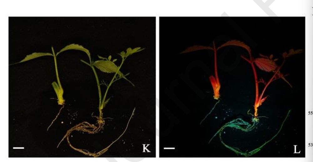
If the prescreen positive rate is clearly higher than expected, check the following items before anything else; the cause can usually be located:
| Source | Appearance | Handling |
|---|---|---|
| Autofluorescence of the substrate | Green over the whole area, blurred boundary, persists when the angle changes | Observe against a dark background and place a negative control in the same substrate |
| Epiphytic algae or colonies on the root surface | Point-like sparkle, scattered distribution | Rinse gently with water and look again; the position shifts |
| Crosstalk from red chlorophyll fluorescence | Green haze around the root | Add a yellow filter and score the tip and mid-section, nothing else |
| Ambient light too strong | Whole root evenly bright, no gradient | Lower ambient illuminance and shorten each exposure |
| Tissue scattering | The wound edge alone is bright, the root body is not | Score at the root tip meristem, not at the wound |
One further point is biological: the paper reports that hairy roots lacking gravitropism and growing upward do not show the RUBY phenotype, with a positive rate of about 70%. In other words, morphologically abnormal roots are inherently unreliable for reporter scoring; even if such roots fluoresce, record and count them separately.
LUYOR-3415RG is the instrument used most often for this kind of prescreen: two wavebands in one unit, with the standard configuration being RB royal blue (440-460 nm, emission 500 nm) and GR green (510-540 nm); RB matches GFP and eGFP, GR matches DsRed, mCherry and tdTomato.
| Item | Specification |
|---|---|
| Excitation wavebands | RB 440-460 nm (emission 500 nm); GR 510-540 nm |
| Light source and power | 12 LEDs across both wavebands, 27 W total |
| Illumination area | Spot diameter about 180 mm at 150 mm from the lamp |
| Power supply | Built-in rechargeable battery, or AC 100-260 V adapter |
| Weight | Net weight about 0.9 kg, supplied in a carrying case |
| Accessories | LUV-30A viewing glasses; LUV-495A 67 mm camera filter (optional) |
One naming point is worth clarifying at the outset: the methods section of the paper writes "LUYOR-3415", which is the series name; the LUYOR-3415RG discussed on this page is the standard configuration within that series with R (royal blue) plus G (green), and it is also the default shipping combination. In other words, the eGFP scoring in the paper falls on the R band; with a different combination (for example R+V for GFP and CFP) the model is still called 3415, but the wavebands differ. List the fluorescent proteins you need to see before ordering, because the combination is fixed at the factory.
The models in the same series fill different roles; choose by sample format:
LUYOR-3415RG two-waveband fluorescent protein excitation source : two wavebands, handheld, suited to rapid prescreening of batches of hairy roots, seedlings and seeds, and usable in greenhouse and field;
LUYOR-3280 : single-waveband portable model, easier on the budget when a project uses a single fluorescent protein (for example a pure GFP system);
LUYOR-3260RB : single-waveband flashlight type, lighter when working with GFP alone or RFP alone;
LUYOR-3421 : benchtop model, suited to continuous viewing of dishes at a fixed position and to cases where images must be archived;
LUYOR-3124RB : large-area suspended type, suited to batch screening of whole seedling trays or larger plants.
If you need both fluorescent proteins and want to carry the light between field and greenhouse, choose the two-waveband 3415RG; for a single-color marker, 3280 or 3260RB is sufficient. The five-waveband code system (R/C/G/V/U) and combination advice are covered in the LUYOR-3415 two-waveband article , which explains in more detail which code matches each fluorescent protein.
It can be seen, but it is not advisable to use that as the basis for scoring. The excitation maximum of eGFP is 488 nm; 365 nm sits on the tail of the absorption curve, where excitation efficiency is low and the signal weak. To see a weak signal you have to dim the surroundings and observe close up, and the result is that background noise cannot be separated from a faint true positive, which pushes the false positive rate higher still.
No. The output of a high-power LED is enough to excite distinguishable fluorescence under ordinary room lighting; move the samples to a light-shielded bench and switch off direct lamps. What really needs controlling is the stability of ambient light, not complete darkness - otherwise the baseline differs from one scoring session to the next.
Two lessons: work in batches by line, take one box at a time and mark each root immediately after scoring so batches do not get mixed; and photograph the position of positive roots through the camera filter so you can sample from the image afterwards, which is more comfortable than cutting on the spot. A benchtop model suits continuous viewing at a fixed position, a handheld model suits screening while moving along the bench.
Yes. The 38.64% false positive rate reported in this paper is the reason. The value of a fluorescence prescreen is to narrow the candidates from hundreds to dozens and reduce the PCR workload, not to replace PCR. The same applies to the RUBY system, where the paper explicitly recommends combining it with molecular verification.
Three causes are common: the root segment was taken but contains no transgenic cells (chimerism or surface attachment), the vector is expressed transiently in the tissue without integration, and the sampled site is too far from the growing point. Molecular results alone can exclude chimerism and transient expression; the third can be reduced by consistently taking the root tip segment.
3415 is the series name; 3415RG is the standard configuration within it with R (royal blue 440-460 nm) plus G (green 510-540 nm), and it is the default shipping combination. The eGFP scoring in the paper used the R band. If you need to view both GFP-type and DsRed/mCherry-type proteins, choose 3415RG; for a single-color marker you can choose the single-waveband LUYOR-3280 . The two wavebands are fixed at the factory, so confirm the fluorescent protein type before ordering.
Yes. Lignin, phenolic compounds and residual chlorophyll in roots can all emit yellow-green background light under blue excitation, and high-polyphenol material such as Rubus is more pronounced. There are three ways to tell them apart. Look at signal morphology: transgenic fluorescence is usually distributed evenly along the root with clear boundaries, whereas autofluorescence tends to be patchy or concentrated at wounds and old root segments. Set wild-type root controls and view them side by side at the same waveband and distance, comparing brightness directly. And cross-check by switching wavebands - eGFP is bright in the R band and should not be equally bright in the GR band, and bright spots that appear just as the waveband changes should be treated with caution.
LUYOR-3415RG two-waveband fluorescent protein excitation source : RB and GR wavebands, 0.9 kg handheld, no darkroom needed;
LUYOR-3280 single-waveband fluorescent protein excitation source : the portable choice for single-color marker systems;
LUYOR-3260RB fluorescent protein excitation source : single-waveband flashlight type, for laboratories working with a single fluorescent marker;
Hairy root transformation of mature macadamia trees: 3415RG screening of fluorescent reporters : how to score eGFP and DsRed2 together in a woody in planta system;
Verification of pea PsLykX gene editing with hairy root fluorescence screening : a comparable record of work in a legume hairy root system;
eGFP colony fluorescence counting in a 2025 Nature alfalfa study : fluorescence counting from colonies through to root systems;
Positive screening of soybean hairy roots: LUYOR-3415RG in GmSIN1 salt tolerance work : another operational record from a similar hairy root system;
The LUYOR-3415 two-waveband approach: high-throughput prescreening of living transgenics : the five-waveband code system and combination selection;
LUYOR-3415RG application cases: six fluorescence viewing scenarios : how scoring differs from seeds and leaves to roots;
Choosing between a UV crosslinker and a fluorescent protein excitation source : how the two kinds of UV tool divide the work, and common misuse.
The real value of this Rubus system is that it lowers the entry barrier from "do you have a tissue culture platform" to "do you have a tray of moist vermiculite and a lamp that can show fluorescence". But once the barrier falls, rigor in scoring matters more, not less: hairy roots all look alike and fluorescence can mislead, and the 38.64% false positive rate is a reminder that visual screening is a filter, not a conclusion. Choose the right waveband, run enough controls and keep molecular evidence for every positive root, and the time saved is genuinely worth having. If you are setting up a hairy root transformation workflow, send us the species, explant type and batch size and we can look at how to configure the fluorescence viewing step, or leave a message through contact us .
This article belongs to the laboratory research techniques section.
Other sections on the same product line include GFP fluorescence flashlights (models grouped by GFP/RFP/DsRed waveband, handheld and benchtop), semiconductor particle inspection (grazing illumination for location, fluorescence for organic material, counting for concentration - three complementary approaches), and life science instruments (the entry point for fluorescence imaging, UV crosslinking, UV transmission and cell irradiation).
Models mentioned in this article: LUYOR-3415RG, LUYOR-3280, LUYOR-3260RB. Ranges, accuracy and accessory lists for each model are given on the product pages.

Email: info@xinysun.com

Tel: 131-2093-6518

Address: 1525 Minqiang Road, Bldg 26, Songjiang, Shanghai

Copyright 2026 Xinysun Instrument (Shanghai) Co., Ltd. All rights reserved.
ICP:ICP-2026046447




 Industrial Test Instruments


