
Products
PRODUCTS Breadcrumb: Home > Products > Life Science Instruments > UV Transilluminators
 Overview
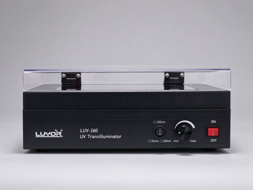
LUV-260 UV transilluminator with 8 x 8W tubes, 260x210mm viewing area, 254, 302 or 365 nm single or dual wavelength, fixed or adjustable intensity.
The LUV-260 series UV transilluminator is used to view and excise nucleic acid (DNA/RNA) gel electrophoresis results. The illuminated area is 260×210mm, so a standard agarose gel lies flat without being moved repeatedly. Eight 8W UV tubes behind special UV filter glass give even brightness across the viewing area, so faint bands can still be captured. A transparent UV shield adjusts to the operator's viewing angle and locks in place, blocking direct UV during viewing, photography and excision.
Single or dual wavelength, fixed or adjustable intensity, and 254/302/365nm - these combinations are set by the model. Choose by whether the main job is viewing gels or excising bands; that is more useful than comparing specifications.
More wavelengths is not automatically better; the band has to match the dye and the purpose:
Excites the EB/DNA complex to fluoresce; bands are bright with high contrast, which makes it the usual band for viewing and photographic records, with good sensitivity.
Lower energy and less damage to DNA, suitable for preparative viewing and gel excision: when the recovered fragment will be used for ligation or transformation, long-wave is the safer choice.
Higher energy, for special viewing that needs short-wave excitation; sample damage and operator protection requirements are correspondingly higher.
A common routine is 302nm to look and 365nm to cut: confirm band position and brightness at mid-range, then switch to long-wave to excise. That workflow needs a dual-wavelength model to stay on one unit; single-wavelength models are chosen for one main purpose.
LUV-260 is the standard fixed-intensity model; when brightness needs to be trimmed to dye concentration, choose the adjustable version (LUV-260A). Dual-wavelength models use four tubes per band and the two bands can be lit separately, so no tube changes are needed when switching.
The transparent shield adjusts to any angle for the viewing position and locks there, blocking UV without blocking the view, so long excision sessions are less tiring.
Transmits the target wavelength and suppresses stray light; sensitivity reaches the ng level, so trace DNA bands are not missed.
Eight 8W tubes behind the filter glass give even brightness across the 260×210mm area, with no dark corners in photos.
The sealed bench surface keeps gel residue from seeping in, protecting the tubes and electronics and extending service life.
A built-in fan keeps heat moving, limits surface temperature rise and reduces the effect of long exposure on gel and sample.
Six wavelength combinations cover viewing, photography and excision; choose by budget and purpose.
Adjust and lock the shield for your viewing height before placing the gel. If the angle is wrong you will lean in unconsciously, and UV exposure rises with it.
302nm for viewing, 365nm for excision. On a dual-wavelength model, locate bands at mid-range first, then switch to long-wave to cut.
Cut decisively once the band is located and avoid leaving the sample under UV; prolonged exposure increases DNA damage and lowers the usability of the recovered fragment.
Fix camera exposure and white balance and use one set of settings for a batch, so band brightness is comparable and later quantification is not distorted.
Wipe off buffer and gel debris promptly and keep the filter glass clear; dust or condensation on the glass directly lowers sensitivity.
Mainly the intensity control: the LUV-260 is fixed, the LUV-260A is adjustable. Fixed suits routine viewing with a stable workflow; if sample concentrations vary widely and exposure must follow band brightness, the adjustable version is handier. Both have a 260×210mm illuminated area and the same wavelength options.
Dual-wavelength models use four tubes per band, and either band can be lit alone or combined as needed. In practice they are usually used in turn: mid-range for viewing, long-wave for excision, which saves tube life and reduces unnecessary light exposure of the sample.
302nm has high excitation efficiency and brighter bands, but also higher photon energy; prolonged exposure increases DNA damage (pyrimidine dimers, for example) and lowers the ligation and transformation efficiency of the recovered fragment. 365nm is slightly dimmer but gentler on the sample, which suits excision where the fragment will be used afterward.
Check three things first: dust, water marks or gel residue on the filter glass; whether the tubes are visibly aged (blackened ends, uneven brightness after ignition); and whether staining and destaining were complete. The first two recover with cleaning or tube replacement; the third points to the staining workflow.
The unit includes a transparent UV shield; keep it in the viewing position during normal viewing and excision. For long close work or photography, add a UV face shield and gloves, and keep the room ventilated: UV exposure generates ozone, especially in small closed spaces.
UV transilluminators branch by dye system and working method; the other two models in this section are worth comparing:
Selection table for the six wavelength combinations of the LUV-260 series
Comparison of 302nm and 365nm for viewing and excision
Routine maintenance and tube replacement interval recommendations
Reference exposure settings for gel viewing and photography
Complete unit and documentation, ready to use on arrival
Advice on wavelength choice, dye matching and operating practice
Long-term supply of UV tubes, filter glass and other consumables and spares
Demo units and on-site technical support in Shanghai, Jiangsu and Zhejiang
Send inquiry

Email: info@xinysun.com

Tel: 131-2093-6518

Address: 1525 Minqiang Road, Bldg 26, Songjiang, Shanghai

Copyright 2026 Xinysun Instrument (Shanghai) Co., Ltd. All rights reserved.
ICP:ICP-2026046447




 Products
 Applications
 Previous:
 Back to list

 Industrial Test Instruments


