
Products
PRODUCTS Breadcrumb: Home > Products > Industrial Test Instruments > Biofilm Detection Lights
Authorized distributor for LUYOR (USA). UV-A fluorescence biofilm detection lamps for food and beverage CIP/COP validation and pharma GMP cleaning validation: under UVA, green fluorescence indicates active bacteria and biofilm while blue indicates dust and fibers; dormant and VBNC microbes can be detected. Covers equipment inner walls, filling valves, conveyors and tank level lines. In stock in Shanghai with compliance solutions.
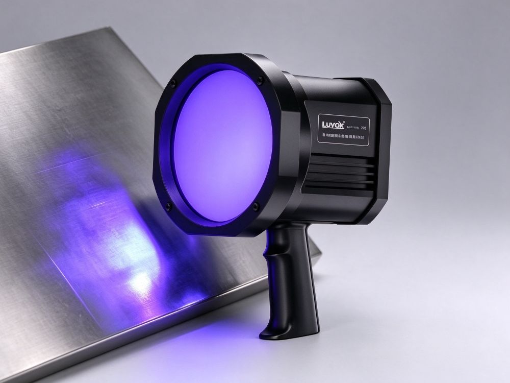
 Model: LUYOR-339

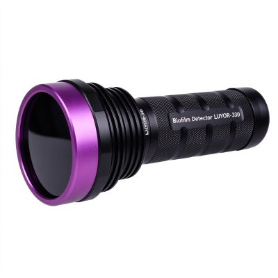
Model: LUYOR-330
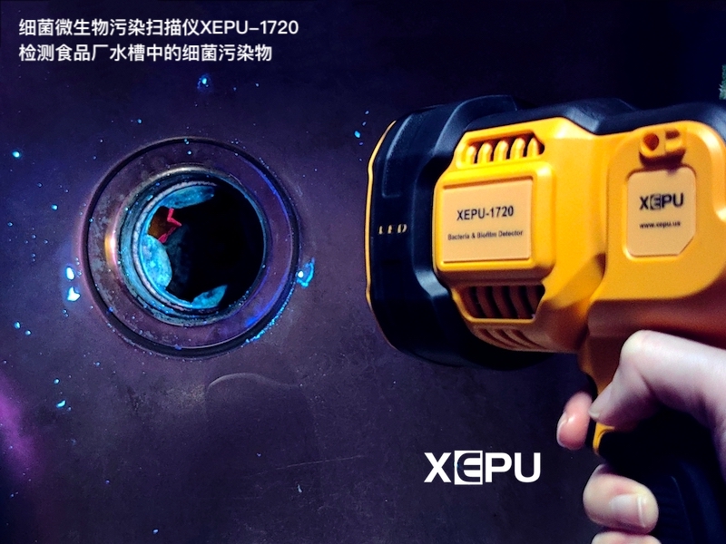
Model: XEPU-1720
The real challenge in cleaning validation is not detection itself but coverage: a workshop may have hundreds of sampling points while swabs can test only a small fraction. The value of a biofilm detection light is that it scans the entire surface and identifies the locations that should be sampled. Choose based on three things: whether it can distinguish biofilm from inanimate debris by fluorescence color, how much area one patrol can cover, and whether it can produce photographic evidence. What it replaces is blind spot-checking — not ATP sampling or laboratory confirmation.
| Model | Brand | Interpretation and Light Source | Runtime / Power | Documentation and Protection |
|---|---|---|---|---|
| LUYOR-330 | LUYOR (USA) | UV-A fluorescence, visible within seconds | Portable battery power | No reagents or consumables, flashlight form |
| LUYOR-339 | LUYOR (USA) | UV-A, green = biofilm / blue = dust | Approx. 5 hours | Green/blue differentiation, detects bacteria in the VBNC state |
| XEPU-1720 | XEPU | UV fluorescence scanning, results within seconds | 2×XP-75 alternating, approx. 2 hours | Non-contact, 1.5kg total weight |
The comparisons above are compiled from publicly available parameters on the individual product pages. For exact configuration, pricing and lead time, please refer to the corresponding product page or call the number at the bottom of this page. Xinysun Instruments (Shanghai) Co., Ltd. supplies LUYOR and XEPU instruments for industrial inspection, UV curing and speed measurement.
A biofilm is the extracellular polymeric substance (EPS) secreted by bacteria after they attach to a surface, composed mainly of polysaccharides and proteins. Under UV-A these substances fluoresce green, while inanimate debris such as dust and fibers usually fluoresces blue. That is exactly what a detection light does: sweep it across a surface in a dark environment and the spots that should be sampled light up on their own.
Its value lies in coverage. A workshop may have hundreds of points while swabs can test only a tiny fraction; sweeping the light over the entire surface and then focusing sampling resources on the spots that actually fluoresce turns random spot-checking into targeted sampling. What it replaces is blind spot-checking — not ATP sampling or laboratory confirmation.
1. Timing: Inspect after cleaning and disinfection are complete and the surface is dry. Water or foam causes fluorescence to spread and distorts interpretation.
2. Environment: Turn off the lights where possible or use a hood — the darker the ambient visible light, the easier it is to see weak fluorescence. If you cannot fully block the light, observe from close range at eye level.
3. Scanning: Keep the lamp head 10-30cm from the surface and sweep slowly at a constant speed, overlapping one third of the beam width to avoid missed or duplicated areas.
4. Interpretation: Look at color first — green usually indicates protein-containing biofilm and active bacteria, blue usually dust and fibers. Then look at distribution — patches appearing along welds, seals and corners are more suspicious.
5. Documentation and sampling: Photograph and number suspicious spots, take targeted swabs for ATP or plate confirmation, and close the loop of detect → sample → verify → recheck.
The main difference is whether you can distinguish biofilm from dust. A model that only shows bright versus dark can easily mistake dust for contamination, leading to unnecessary cleaning shutdowns.
| Type | Capability | Suitable Applications | Limitations |
|---|---|---|---|
| UV-A single wavelength | Rapid visualization of residue distribution | Routine patrols, post-cleaning acceptance | Cannot distinguish biofilm from dust |
| UV-A two-color discrimination | Green = biofilm, blue = dust | Cleaning validation, audit documentation | Still requires a dark environment and experienced interpretation |
| Multi-band alternation | Four alternating excitation bands for stronger contrast | Food, beverage and pharmaceutical pipelines and tanks | Heavier equipment and higher training cost |
| With imaging / recording | On-screen imaging and photographic records | Applications requiring reports and traceability | Higher price; keep the lens clean |
- Food and beverage: Tanks, pipe welds, filling heads, conveyor belts and returnable crates.
- Pharmaceuticals and cosmetics: Mixing tanks, valves, seals, cleanroom drains and wall corners.
- Water treatment and cooling systems: Tank inner walls, pipe elbows and scrubber packing.
- Medical and laboratory: Instrument lumens after cleaning, sinks and biosafety cabinet work surfaces.
- Marine and storage tanks: Ballast water tanks, tank bottom sludge and microbiologically influenced corrosion of inner walls.
- Cleaning validation training: Let operators see for themselves the gap between looks clean and actually has residue.
- Selection and Brands: How to Choose a Biofilm Detection Light? Answers to 10 Common Questions (Authorized LUYOR Distributor)
- Operation and Interpretation: What Is Biofilm Monitoring? Six Detection Methods and On-Site Fluorescence Screening, Biofilm Detection in Pharmaceutical Cleanrooms: How to Find Contamination You Cannot See, Biofilm Imaging Methods: UV Lamp Fluorescence Detection with Photographic Comparison, How to Perform Biofilm Detection in Food Plants: EPA Methods and Multi-Wavelength UV Equipment Selection
No. Fluorescence reveals residual protein and microbial extracellular material and is a screening tool for quickly locating where to sample; the regulatory criterion remains ATP sampling, plate counts or laboratory confirmation. The correct procedure is to sweep the light over the whole surface, mark the green fluorescent spots, then take targeted samples at those spots, spending consumables where there is a real problem.
Under UV-A, green fluorescence usually comes from extracellular-protein-containing contamination such as biofilm and active bacteria, which cannot be removed by simple wiping; blue fluorescence usually comes from inanimate debris such as dust and fibers, which wipes away easily. The value of distinguishing them is avoiding two kinds of error: neither shutting down for a major clean because of loose dust, nor simply wiping over a real biofilm as if the surface were a bit dirty.
Yes. Under stress such as nutrient deprivation or disinfectant challenge, bacteria can enter a dormant, stressed or even VBNC state: they are alive but do not form colonies on culture media, so the report reads not detected. Fluorescence is based on the fluorescent reaction of extracellular proteins and is independent of the organism's current culturability, so it fills this blind spot of culture methods.
Calculate by area covered rather than by room: keep the lamp head 10-30cm from the surface, move at a constant speed with one-third overlap, and once practiced you can cover roughly 2-4 m² per minute. For a 500 m² workshop, focusing on critical areas (equipment surfaces, welds, drains) typically completes a round in 1-2 hours, which is more realistic than scanning every square meter.
It does not have to be a darkroom, but ambient light should be minimized. The stronger the visible light, the more easily weak fluorescence is washed out. If the workshop lights cannot be turned off, use a hood, observe close up inside equipment or in a corner, or choose a model with a filtered viewing window. In fully lit conditions, detection sensitivity drops markedly.
Fluorescence first, ATP second. Fluorescence finds the locations: sweep the entire surface and mark suspicious spots; ATP provides the numbers: take targeted samples at those spots. Reversing the order degenerates into random spot-checking and leaves the coverage problem unsolved. If the two disagree, the laboratory plate count or species identification takes precedence.
A recheck immediately after disinfection only proves that the surface was clean right after disinfection, which is of limited value. A more practical approach uses three time points: after cleaning (to verify cleaning effectiveness), after disinfection (to verify disinfection coverage), and after 4-8 hours of production (to check whether biofilm rapidly re-establishes). Only by looking at the three results together can you tell whether the cleaning process is truly effective.

Email: info@xinysun.com

Tel: 131-2093-6518

Address: 1525 Minqiang Road, Bldg 26, Songjiang, Shanghai

Copyright 2026 Xinysun Instrument (Shanghai) Co., Ltd. All rights reserved.
ICP:ICP-2026046447




 Products
 Applications
 Industrial Test Instruments


