
Products
PRODUCTS Breadcrumb: Home > Products > Industrial Test Instruments > Biofilm Detection Lights
 Overview
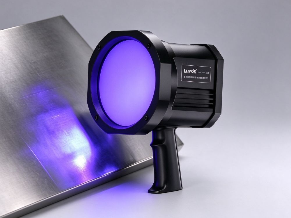
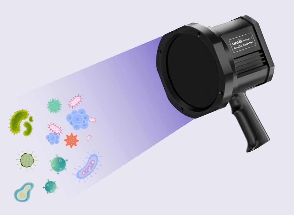
LUYOR-339 is a cordless UVA inspection torch for cleaning validation, with green and blue fluorescence reading and about 5 hours of runtime per charge.
The difficulty in cleaning validation is not the measurement itself. It is coverage. A plant may hold hundreds of points: tank inner walls, conveyor belts, filling valves, pipe joints, gaskets, the floor around drains, laminar-flow grille faces. Swab sampling required by regulation can cover a small fraction of them, and whether the rest is clean is judged by how it looks.
Biofilm grows where it is hard to see and hard to brush. When the few sampled points happen to pass, a biofilm site hidden in a dead corner keeps releasing contamination into the next production run, or the next batch, until a finished-product test fails. By then the cost of tracing the cause is high.
LUYOR-339 addresses this coverage problem. It does not sample; it scans the whole surface. The higher-power source widens the irradiated footprint, so the same inspection time covers far more area. Around 5 hours of continuous runtime means a full line or a cleanroom zone can be walked in one pass, without a return to the charger. Residue shows as fluorescence in a darkened room, so which point is affected and how the problem is distributed are visible at a glance.
This is a capability of the LUYOR-339 that is easy to overlook. Under UVA it does more than light up soil: it separates contamination by type through colour, so the operator can decide on the spot which cleaning route applies:
| Fluorescence colour | What it indicates | After wiping with a glove | Action required |
|---|---|---|---|
| Green fluorescence | Soil that contains extracellular protein - biofilm and active bacteria | Does not wipe off; a simple wipe will not remove it | Treat as a contaminated point, run the cleaning and sanitising procedure, and confirm that point with ATP or laboratory testing where needed |
| Blue fluorescence | Dust, fibers, and other inanimate debris | Wipes away at once | Routine wiping is sufficient; no microbiological risk |
The practical value of this distinction is that it prevents two kinds of misjudgement: a film of dust is not treated as microbiological contamination that stops the line for cleaning, and a genuine biofilm is not dismissed as a slightly dirty surface and wiped over. For audits, the green/blue split also strengthens the record - a photograph of green fluorescence is direct visual evidence that soil is present.
Plate culture rests on one assumption: the organism must grow on the medium. Under nutrient limitation, disinfectant stress, or temperature swings, bacteria enter a dormant or stressed state, or shift into VBNC (viable but non-culturable) form - they are alive, they metabolise, and they can revive and cause illness when conditions allow, but they do not form colonies, so the report reads "not detected".
LUYOR-339 does not depend on growth. It reads the fluorescence of extracellular protein in the soil under UVA. As long as the protein is present, it shows, whatever the current culturability of the cells. That is why it works as well on dormant cells, stressed cells, and VBNC organisms.
For cleaning validation the difference is practical: a negative culture does not mean the surface is clean. Running a fluorescence survey as a pre-screen ahead of culture lowers the chance that a missed site appears in the next batch.
LUYOR-339 biofilm detection lamp body - industrial housing for food, beverage, pharmaceutical, and healthcare sites
UVA safety glasses - UV protection for long sessions
Wide-voltage wall charger - suits different supply conditions
Aluminum hard carry case - transport between areas and equipment storage
Cordless battery power, with around 5 hours of continuous runtime
Efficient cooling keeps the head from overheating during extended operation
Light and portable, and the case eases moves between plants and points
Green/blue reading needs no extra equipment
| Setting | What it does there | Areas to watch |
|---|---|---|
| Food, beverage, and dairy processing | Verifies hygiene on equipment and pipe surfaces after CIP / COP | Tank walls, pipe joints, valves, gaskets, conveyors, filling heads |
| Pharmaceutical and biotech plants | Cleaning validation of production equipment and the production environment | Buffer tanks, pipe connections, laminar-flow return grilles, walls and floors |
| Hospitals and dental clinics | Reduces cross-contamination risk in aseptic areas | Instrument lumens, tray corners, treatment units, interiors of washer-disinfectors |
| Restart and changeover checks | Confirms cleanliness before production resumes and avoids batch deviation | Whole line in one sweep, with attention to residue from the last shutdown |
| Hygiene audits and quality control | Provides point records and images for audit preparation | Work through the audit checklist and record green fluorescent points |
| Other sectors | Identifies surfaces that fall short of hygiene standards | Commercial kitchens, hotels and resorts, breweries, cleanrooms, water treatment and cooling towers, cruise and marine, research laboratories |
Runs routine environmental monitoring and records survey results by point.
Works alongside conventional testing to close the blind spots of culture methods.
Audits cleaning performance and defines the scope of rework quickly.
Manages risk proactively and prepares image evidence for audits.
Divide the scope into zones by room or equipment and plan a route with no backtracking. The usual failure in a large survey is a missed zone, so fix the route before the sweep begins.
Lower the site lighting to raise fluorescence contrast. A cleaned surface needs to dry fully, because a water film reflects light and disturbs the reading.
Sweep at a steady pace along the planned route. Mark green fluorescent areas as contaminated points and treat blue areas as inanimate debris. Where green and blue mix, judge by the green portion.
Mark each green fluorescent point and photograph it, recording position and severity. The images support rework and become part of the cleaning validation file.
Take an ATP swab or a laboratory sample at marked points, so consumables are not spent on areas that are clean. After corrective work, re-check along the same route and compare the fluorescence to confirm the result.
How the three models in the LUYOR biofilm detection range differ:
| Model | Form factor | Key characteristics | Typical application |
|---|---|---|---|
| LUYOR-339 | Handheld detection lamp (higher-power version) | Higher power and a wider irradiated footprint, around 5 hours of runtime, green/blue fluorescence reading | Fast sweeps of large bench, wall, and tank exteriors, whole-zone surveys, long continuous sessions |
| LUYOR-330 | Portable torch format | Compact body with a large head face, easy to reach into tight spaces | Pipe interiors, equipment gaps, complex parts, surveys that need flexible positioning |
| LUYOR-0904 | Canopy lamp for visible-foreign-matter inspection | Inspection lighting configured to the standard | Visible-foreign-matter inspection stations for injectables |
The choice comes down to one sentence: take the 339 when area coverage and session length are the constraint, and take the 330 when access to narrow spaces is the constraint. The detection principle is the same. Many customers keep one of each by zone, the 339 for the broad sweep and the 330 for complex structures.
The attributes below can be confirmed and used for an initial selection:
| Product type | Handheld biofilm detection lamp (non-contact fluorescence detector) |
| Light source | Long-wave ultraviolet (UVA) |
| Power | Cordless battery; around 5 hours of continuous runtime |
| Thermal design | Efficient cooling for extended continuous operation |
| Detection method | Non-contact visual fluorescence reading; no liquids, dyes, or reagents |
| Reading basis | Green fluorescence = biofilm / active bacteria; blue fluorescence = dust, fibers, and other inanimate debris |
| Detectable states | Dormant cells, stressed cells, and VBNC (viable but non-culturable) organisms |
| Construction | Industrial housing for food, beverage, pharmaceutical, and healthcare sites |
| Standard kit | Lamp body, UVA safety glasses, wide-voltage wall charger, aluminum hard carry case |
| Sectors | Food, beverage, and dairy; pharmaceutical and biotech; hospitals and dental; cleaning validation and hygiene audits |
Ultraviolet output falls gradually with operating hours. As irradiance drops, fluorescence contrast weakens and a false negative that looks normal becomes more likely. Check head output periodically with a UV radiometer, compare it against earlier readings, and include the lamp in the equipment inspection and calibration schedule.
Under UVA, soil that contains extracellular protein fluoresces green and indicates biofilm and active bacteria; blue fluorescence indicates dust, fibers, and other inanimate debris. Wipe with a gloved hand to confirm: a blue area usually clears at once, while active contamination in a green area does not come off with a simple wipe and needs the cleaning and sanitising procedure. The reading separates dust from active growth on the spot.
The reading is based on the fluorescence of extracellular protein under UVA and does not depend on growth on a medium. Dormant, stressed, and VBNC (viable but non-culturable) organisms therefore show as well, and those are the states that plate culture tends to report as negative, which is how a missed site arises.
The rated continuous runtime is around 5 hours, and efficient cooling supports extended operation. At the pace of a plant-wide sweep that usually covers a full zone survey or the validation of several lines without a return to the charger. Actual time depends on ambient temperature and how long the lamp stays on.
The LUYOR-339 lamp body, UVA safety glasses, a wide-voltage wall charger, and an aluminum hard carry case. The glasses and the case ship with the lamp, so the kit is ready for site use out of the box.
The 339 is the higher-power version with a wider irradiated footprint. It suits fast sweeps of large benches, walls, and tank exteriors, and whole-zone surveys that run for hours. The 330 is a compact torch that reaches pipe interiors and equipment gaps. Choose the 339 when coverage and session length are the constraint, and the 330 when access is the constraint; the two can also be paired by zone.
No. It is an aid to a compliance programme. Its value is rapid, non-contact location of contaminated areas, so that swabs, ATP tests, and cultures go to the points that need them, which reduces consumable use, shortens downtime, and provides point records and images for audits. Validation data required by regulation still comes from the standard test methods.
There are two common patterns. For post-cleaning verification, scan tank walls, pipe joints, valves, gaskets, and dead corners once CIP/COP or manual cleaning is complete and the surface is dry; concentrated green fluorescence marks the points that need rework. For pre-start checks, sweep the whole line before a restart or changeover and confirm the state before the line runs, which avoids batch deviation from residue. Cleanrooms follow the same logic for equipment and environment, with points and images filed in the cleaning validation record.
UV output falls gradually with operating hours. Check head irradiance periodically with a UV radiometer and compare it against the factory record or earlier readings.
Xinyisheng Instruments (Shanghai) keeps LUYOR instruments in stock and can arrange an on-site demonstration and a trial in your own process. Call 131-2093-6518, or leave your sector, application, and contact details in the form at the bottom of this page.
Zone the area, then plan the route. Divide the scope into zones by room or equipment, plan a route with no backtracking, and work through the zones in order. The usual failure in a large survey is a missed zone, so fix the route before starting and record completion by zone so that a re-check can follow the same path.
Yes. UV output falls gradually with operating hours, and as irradiance drops the fluorescence contrast weakens, which makes a false negative that looks normal more likely. Check head output periodically with a UV radiometer, compare it against earlier readings, and include the lamp in the equipment inspection and calibration schedule.
The detection principle is the same; the difference is form factor and coverage. Choose the 339 when coverage and session length are the constraint: higher power, a wider irradiated footprint, and around 5 hours of runtime suit fast sweeps of large benches and tank exteriors and whole-zone surveys. Choose the 330 when access is the constraint: the compact body reaches pipes and equipment gaps. Many customers keep one of each by zone, the 339 for the broad sweep and the 330 for complex structures.
LUYOR-339 specification sheet and product manual
Fluorescence survey proposal for cleaning validation
LUYOR biofilm detection range selection table
Stock in Shanghai, with on-site demonstration and trial evaluation
Selection advice, operator training, and survey procedure review
Manufacturer warranty, with long-term supply of parts and protective consumables
Related reading:Biofilm in food and beverage: rapid detection for safer production | Beyond the surface: advances in bacteria and biofilm detection | See also:LUYOR-330 portable biofilm detection torch
Send inquiry

Email: info@xinysun.com

Tel: 131-2093-6518

Address: 1525 Minqiang Road, Bldg 26, Songjiang, Shanghai

Copyright 2026 Xinysun Instrument (Shanghai) Co., Ltd. All rights reserved.
ICP:ICP-2026046447




 Products
 Applications
 Previous:
 Back to list

 Industrial Test Instruments


